29
Interventional Management of Gastrointestinal Strictures
Strictures of the gastrointestinal (GI) tract have many causes, including malignant, congenital, benign, and postsurgical lesions. Although these strictures can be difficult to manage, surgery has been the standard method of treatment; however, endoscopic and interventional radiologic treatments are rapidly proving to be effective methods in caring for these patients. In the upper GI tract, esophageal stenoses are the most common lesions, followed by gastric and duodenal lesions; newer techniques now allow interventional management of colonic strictures. All these lesions are amenable to stricture dilation.
 Esophageal Strictures
Strictures of the esophagus may be secondary to primary congenital anomaly, caustic ingestion, malignant disorders, esophageal surgery, or esophagitis. Malignant strictures arise directly from a primary esophageal carcinoma or from an adjacent lung or mediastinal malignancy invading the esophagus. In the midesophagus, 90% of the primary esophageal malignancies are due to squamous cell carcinoma. In the distal third of the esophagus, the most common malignancy is gastric adenocarcinoma. Treatment of these malignant disorders has included surgical bypass, stent placement, or primary repair. Although 50% of esophageal tumors are deemed resectable, the 5-year survival rate of these patients is only 10%, regardless of the treatment.1,2 The most commonly encountered benign etiology for strictures in adults is reflux esophagitis. Other causes include achalasia, caustic ingestion, postradiation changes, and iatrogenic trauma. Traditional treatment of these benign strictures is repetitive dilation with mercury bougies.
Transluminal dilation with angioplasty balloons has added yet another effective tool to the treatment armamentarium of these stenotic lesions. When used with fluoroscopic and standard guidewire techniques, these balloons have proved safe and effective. The recent availability of expandable metallic stents has added to the interventional radiologists ability to assist in the treatment of these patients, particularly those with malignant disease.
Surgical and endoscopic treatments
Traditional surgical methods for the treatment of esophageal strictures include patching the esophagus in the region of the stricture or bypassing the stricture with colon or jejunum. Although the former method may result in excellent relief of dysphagia and reflux symptoms, about one half of patients undergoing patching may require dilation at a later date. Intestinal interposition operations are effective but also are associated with significant morbidity and mortality and constitute a major surgical procedure. For malignant disease, radiation therapy and chemotherapy have short-lived effects, and although laser treatment is used for exophytic tumors, it must be repeated frequently.
Bougienage has been performed for centuries and involves the use of an instrument with a straight or rounded olive-shaped tip inserted through the stricture, progressively stretching the narrowed segment to achieve the desired lumen. In the past, these instruments could be passed repeatedly by the patient using bougies (such as the Hurst or Maloney dilators); however, the use of endoscopic techniques has encouraged the development of types of bougies that pass over guidewires, reducing the likelihood of perforation. The most popular of these models are the Celestine and Eder-Puestow devices.
Although effective, these devices have one major disadvantage: They provide a significant radial force on the stricture but also exert longitudinal force on the adjacent, normal esophagus. This additional stress can lead to perforation or mucosal tears.3 Angioplasty balloons passed over a wire and placed directly across the lesion may overcome the shortcomings of bougienage. These balloons can be introduced on a small catheter that then can be inflated to a large outer diameter, thus maximizing the radial force on the lesion while reducing the longitudinal force on the adjacent esophagus.
General intubation techniques
Before esophageal dilation, an esophagram is required to help delineate the anatomy of the stricture. Stricture dilation generally does not require analgesics, and sedation with intravenous midazolam hydrochloride (Versed) is usually adequate. Atropine may be helpful if the patient develops signs of increased vagal tone (e.g., bradycardia) or if secretions are copious. The use of topical analgesia such as anesthetic gels or oral sprays are recommended to avoid patient discomfort and to reduce the gag reflex.
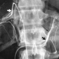
A soft nasogastric tube is passed transorally and placed just above the level of the stricture. The transoral intubation approach usually is employed for esophageal stricture dilation. This transoral route is easier and better tolerated, and it will permit passage of catheters larger than those used in the transnasal approach. Moreover, this route must be used for the placement of stents. The transnasal approach is preferred if a feeding tube is to be placed over the same guidewire. By enlarging the side hole in the nasogastric tube, the rounded tip of the tube can be maintained while still allowing guidewires to be inserted through the tube. After the tube is in position, a small amount of contrast is injected to define the proximal aspect of the stricture. In patients at risk for aspiration, dilute barium or oily contrast may be used.
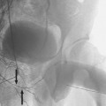
A flexible-tipped standard 0.035-inch guidewire then is passed through either the endhole or sidehole of the nasogastric tube, which then is exchanged for a 5 Fr selective angiographic catheter. Although the stricture often can be traversed using a flexible guidewire, a torque-control guidewire or hydrophilic guidewire may be used to cross the lesion if the stricture is particularly tight or irregular. Great care must be exercised to minimize the risk of esophageal perforation, particularly in patients who have malignant strictures. Once the guidewire and catheter are through the lesion, the guidewire is removed and replaced with a long exchange wire (>
Related posts:


Stay updated, free articles. Join our Telegram channel

Full access? Get Clinical Tree


