Fig. 13.1
(a) 3D apical tumor (arrows) with capsule intact. (b) MRI irregular focal tumor (arrows). (c) DCE-MRI no enhancement is noted
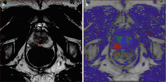
Fig. 13.2
(a) MRI focal midgland tumor right. (b) DCE-MRI homogeneous enhancement
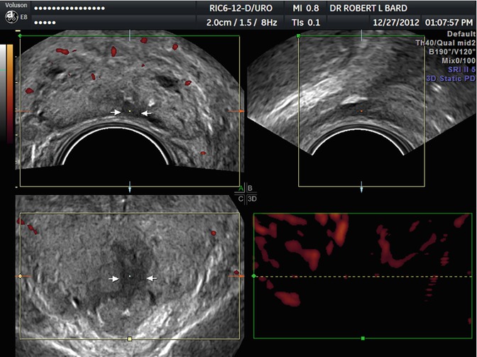
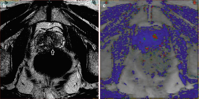
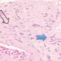
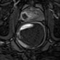
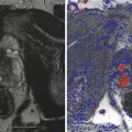
Fig. 13.3
(a) 3D 4 mm avascular apical tumor (arrows). (b) MRI tumor poorly visualized (arrow). (c) DCE-MRI no tumor enhancement
Related posts:

Stay updated, free articles. Join our Telegram channel

Full access? Get Clinical Tree


