SONOGRAM ABBREVIATIONS
IUD Intrauterine device
IUCD Intrauterine contraceptive device
KEY WORDS
Copper T and Copper 7 IUDs. Intrauterine devices (IUDs) containing copper that are still in use.
Lippes Loop. Obsolete but widely used serpentine-shaped IUD constructed with five parallel portions of plastic.
Levonorgestrel. Progesterone-like agent used in modern IUDs that release hormones over long periods.
Mirena Intrauterine System. T-shaped device that slowly releases levonorgestrel over a 5- to 7-year period.
Pelvic Inflammatory Disease (PID). Infection that spreads throughout the pelvis, often caused by gonorrhea. If it is secondary to an IUD, other bacteria are usually found.
Progestasert. T-shaped IUD that releases progesterone. Needs to be replaced annually.
The Clinical Problem
Intrauterine contraceptive devices (IUDs, IUCDs) are a highly effective means of contraception with relatively few systemic side effects. Although many varieties of IUDs have been used, only the most commonly used devices will be discussed here. Less than 1% of women in the United States use these devices; however, 15% to 30% of women use IUDs in Europe and Canada.
Because pelvic inflammatory disease (PID) was a common side effect in women who used IUDs, the devices fell out of favor. Modern IUDs that slowly release progesterone or a progesterone-like substance, levonorgestrel, over a number of years are making a comeback because infection is rare nowadays. Usually inserted to prevent pregnancy, IUDs may also be used to prevent dysfunctional bleeding.
The proper location of an IUD, regardless of type, is in the endometrial cavity at the uterine fundus. The remainder of the device should be above the cervix. A nylon thread, which extends from the uterus into the vagina, is attached to the proximal end of all IUDs. This string should be palpable or visible on pelvic examination. If this string cannot be identified, the patient may be referred for evaluation of a “lost IUD.”
Some patients have no complaint other than a lost string. Others, however, present with cramping, pain, or abnormal bleeding. In either case, the position of the IUD must be demonstrated. If the uterus is empty, the device has been expelled or has perforated the uterus. An IUD outside the uterus is usually not seen with ultrasound because it is surrounded by gut. Although an IUD in the correct location prevents a normal pregnancy effectively, ectopic pregnancies may still occur.
Anatomy
Anatomy of the pelvic area is discussed in Chapter 29.
Lippes Loop
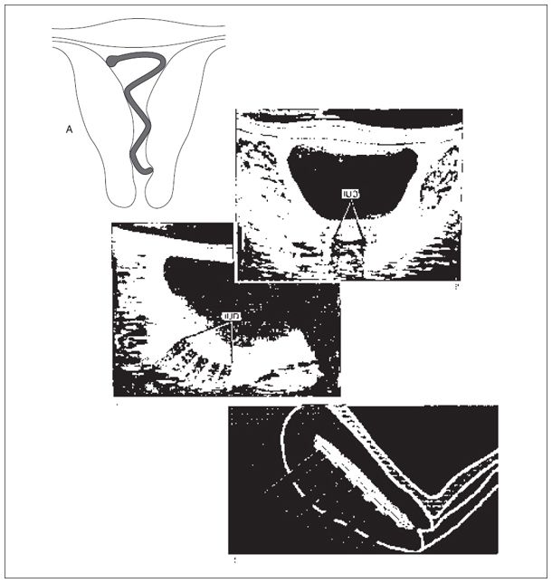
The Lippes loop was the most widely used IUD, and there are still some in use. In a long-axis view, the loop has two to five echogenic components, depending on whether a true long-axis IUD view has been obtained (Fig. 34-1A,B). Transversely, the device is visualized as a single line.
Dalkon Shield
Insertion of the Dalkon shield was suspended some years ago because of a large number of associated infections. There are still a few women using the device. The Dalkon shield is the smallest of the IUDs. On both longitudinal and transverse scans, it appears as two echogenic foci.

Figure 34-1.
Stay updated, free articles. Join our Telegram channel

Full access? Get Clinical Tree


