, Jean-Philippe Bault2, Bernard Benoit3 and Gérard Couly4
(1)
Center of women and fetal imaging, Créteil, France
(2)
Center of fetal imaging Ambroise Paré, Les Mureaux, France
(3)
Princess Grace Hospital, Monaco, France
(4)
Department of maxillo-facial surgery, Necker Hospital, Paris, France
The facial bones, as they appear in a fetal ultrasound image, result from a continuous process of development, regulated and sequenced by genetic and molecular embryology, which progressively constructs the face like epigenesis.
The face, like the entire head, is “prefigured” by prescribed regions that are genetically determined during the gastrula and neurula stages. Via typological deformations, the two-dimensional neural surface transforms symmetrically and acquires a third dimension.
When the dorsal region of the neural grove closes into a tube, the “fourth layer” of migrating neural crest cells invade the future embryonic cephalic pole and provide the cellular filling for the facial prominences.
The face develops thanks to the five founding prominences or swellings of the ectoderm, which grow volumetrically and then fuse by contact and are no longer recognizable as having existed once the face is totally formed. These prominences are:
The frontonasal prominence, which is median, single, symmetrical and different from the other four as it is formed by two left and right halves.
The maxillary prominences, right and left.
The mandibular prominences, right and left (first branchial arches).
1.1 The Key Stages in Craniofacial Development
The prescribed regions of the face and brain are differentiated as early as the gastrula and neurula stages .
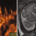
Gastrulation lays out the embryonic body plan for all vertebrates with bilateral symmetry via the formation of the notochord and the mesoderm interposed between the endoderm and the ectoderm (Fig. 1.1) (this beginning explanation of epigenesis or the phenomenology of development by stages replaced the eighteenth century concept of preformation that stemmed from the building of the first microscope, when Leeuwenhoek, its inventor, identified the spermatozoid in 1677).
The future regions of the face are situated in the rostral regions of the gastrula.

Fig. 1.1
Preformationism vs. epigenesis. 1 Homunculus. 2 End of gastrula phase, beginning of neurula stage
Neurulation is a theater of morphogenetic operations that will differentiate the prescribed regions of the head (Fig. 1.2). This is the stage during which the central nervous system, the facial mass and the neck truly begin their morphogenesis.
The neural plate is the two-dimensional cellular surface whose fate mapping demonstrates the closeness and unity of the regions of the brain, the neuro-sensorial receptors, and the face, genetically “printed” on the neural plate.
During neurulation, the topological transformation of the neural plate continues. The face is in the neurula and “grows” in front of it (Fig. 1.3).
The neural plate lengthens and folds, forming a groove with edges rising up and fusing into a tube (Figs. 1.4 and 1.5).
Closure of the neural tube’s rostral opening represents an exceptional topological originality (Fig. 1.6): the anterior-most parts of the neural plate roll up forward, completing a half-circle rotation (180°), flipping these regions into a ventral position (it is an elegant and original solution to the problem of closing a tube that is open in the front (Figs. 1.7 and 1.8).


Fig. 1.2
Map of neural plate regions
Fig. 1.3
The face grows in front of the neural plate
Fig. 1.4
Closure of the neural plate into the neural tube, with neural crest
D 15 – The neural plate
D 18 – Dorsal fusion
NC Neural crest
Neural tube
Fig. 1.5
Topogenesis of the anterior neural plate and beginning of the optic cup.
The optic cups invaginate. Elevated neural ridges move closer together on the dorsal side of the embryo
Frontal view
Fronto-lateral view
Mouse, D8-D8, 5; human, around D22
Fig. 1.6
Overall ventral rotational movement of the rostral extremity of the embryo
1. Rotation–D20
2. Facial cerebral expansion – D30
3. Ventralization – D 40
Fig. 1.7
Optical vesicles and the median closure of the rostral extremity
In this cut, one can observe the links between:
The optical buds, the ectoderm and the mesenchyme
Anterior
Posterior
Mouse D8, 5; Human around D22-24