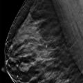
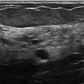
Introduction to Localization and Biopsy Using Tomosynthesis Digital breast tomosynthesis acquires several images of the breast at multiple angles. The individual images are reconstructed into a series of thin, high-resolution slices typically 1-mm thick, which can be viewed like computed tomography (CT) or magnetic resonance (MR) images as a single slice or stacked in dynamic cine mode. The ability to view images as a single slice or in dynamic mode eliminates the effects of overlapping breast tissue, which allows for better visualization and characterization of masses. Conventional two-dimensional (2D) mammography plus digital breast tomosynthesis increases reader performance for identifying masses and architectural distortions over conventional mammography alone. DBT can find lesions not seen on conventional 2D mammography. The addition of DBT reduces callbacks for false-positive lesions and has a higher breast cancer detection rate than conventional mammography alone, specifically in the detection of small invasive, node-negative cancers. Although DBT is excellent at characterization of benign and malignant features, definitive diagnosis is proven by histopathologic analysis. Many small architectural distortions detected by tomosynthesis may not be seen with sonography, thereby negating sonographic biopsy. Prone stereotactic vacuum-assisted biopsy of an architectural distortion is susceptible to targeting error because the imager may not be able to identify the architectural distortion on both stereo pair images. Also, the biopsy window for prone stereotactic biopsies is small, requiring a skilled technologist to properly position the finding within the biopsy window. Tomosynthesis-directed stereotactic biopsy uses the full detector for imaging and biopsy. Additionally, calcifications are often better biopsied with stereotactic biopsy than with ultrasound. The three-dimensional (3D) capability of tomosynthesis allows lesions to be detected at the correct depth. Tomosynthesis-directed stereotactic biopsy has the advantage over prone stereotactic biopsy of calculating the distance from skin to lesion on the orthogonal images and thus facilitating the most efficacious approach for biopsy. Tomosynthesis-directed stereotactic seems better for low-contrast lesions such as uncalcified masses and architectural distortions. Tomosynthesis-directed biopsy can be performed quickly and safely without major complications. One limitation is that needle adverse patients may have difficulty seeing the biopsy needle. One solution is an eye mask or using a prone stereotactic table.
Related posts:


Stay updated, free articles. Join our Telegram channel

Full access? Get Clinical Tree


