CHAPTER 19 Introduction to the reporting of gastrointestinal (GI) radiological procedures


Introduction
The reporting of radiological (medical imaging) procedures has traditionally been performed by radiologists (specialist medical doctors). However, initially in medical ultrasound (US), due to increasing workload and a shortage of radiologists beginning in the 1970s, there has been a gradual shift within the UK to reporting duties being undertaken by allied healthcare practitioners, predominantly radiographers (Fernando, 1999).
The precedent from US imaging was expanded into Accident and Emergency (A&E) radiographic imaging, initially with an abnormality flagging system, often called the ‘red dot’ system (Berman et al., 1985; Nuttall, 1995). This subsequently developed into reporting of a wider range of plain radiographs by radiographers (Quick, 1993; Robinson, 1996a; Robinson et al., 1999a) and from there expanded into most other subspecialties of medical imaging (Culpan, 2006).
Although there is no specific requirement that the delegation of medical image reporting be to a specific professional group (General Medical Council, 2001), the delegation process should be robust and take into account the skills base of those to whom the task is delegated.
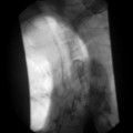
In the field of GI imaging, the first area of delegated practice to be undertaken (and subsequently reported) by radiographers was the double contrast barium enema (DCBE) (Mannion et al., 1995). Radiographer reporting of DCBE was shown to be of similar accuracy to radiologist reporting (Law et al., 1999; Culpan et al., 2002, 2003; Booth and Mannion, 2005).
Reporting standards
Standards of reporting are important, not least because cases may be tested in a Court of Law. As such, the standards achieved by the healthcare professional undertaking the delegated reporting role must at least match the standards of the professional group which had previously performed the role, namely the clinical radiologists (Dimond, 2002, 366-7). The RCR produced standards for reporting for radiologists (The Royal College of Radiologists, 2006) which consequently must also apply to all others undertaking the reporting role.
Practical skills
Observation and interpretation
Digital format has several advantages (e.g. the potential for manipulation of the image after it has been captured); however, the uninitiated may simulate or mask/obscure pathology by incorrect manipulation of images. Transfer of images in digital format is also simplified; no longer does the reporting professional have to wait for ‘hard copy’ images or carry around stacks of photographic film. Electronic digital image files can now be transferred to image reporting stations within (and outside) the healthcare facility in which they are acquired.
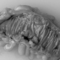
Although pattern recognition has its uses, there are potential downfalls to relying on the process. For instance, if a pattern demonstrated is not one that the viewer has seen before, or is not one they are completely familiar with, it may not be recognized. In such cases, even quite obvious abnormalities may be ignored as the individual is only looking for ‘what they know’. To overcome this limitation, the trainee reporting practitioner is taught to review medical images systematically, interrogating the whole image sequentially, looking at all parts of an image and all images within each particular examination or study. This may be quite challenging since with multidetector CT examinations (MDCT) and multisequence MR studies, there may be 300–500 individual images in a single study (Fishman, no date). When, therefore, it is not possible to scrutinize each image in detail, pattern recognition remains the primary method of review. This process may be facilitated by computer-assisted detection (CAD) software, application specific programs being commonplace for virtual colonoscopy (VC) for instance, to highlight potential pathology.
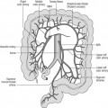
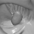
It is also important that personnel involved in image interpretation and reporting understand the clinical information provided by the referring clinician (The Royal College of Radiologists, 2006). In the UK, there is a legal requirement that this is sufficient to enable the radiologist or radiographer to ‘justify’ the examination with respect to the ionizing radiations (medical imaging) regulations (IR(ME)R 2000) (Department of Health, 2000). Clinical information in the referral should convey any relevant signs and symptoms elicited from the patient’s medical history, physical examination and laboratory tests and should give a provisional diagnosis or suggest differentials. The referral is often expressed in terms of a ‘clinical question’, for instance, a patient with unexplained iron deficiency anemia may be referred for a range of diagnostic tests such as initial esophago-gastroduodenoscopy (OGD) and subsequent DCBE or colonoscopy to confirm or refute the presence of a colonic source of bleeding such as a cecal tumor.
Reporting style and structure
Most medical imaging reporters use a free text type report, although alternatives include a pre-defined proforma style, where specific questions are answered or pre-coded standard phrases selected (Naik et al., 2001). Each style has its own strengths but the recommended structure expected of medical imaging reports is defined by the The Royal College of Radiologists (2006). Initially, a report should contain patient identifying information to ensure that a specific report can be related to a specific patient on a specific occasion, as misidentification can have significant, potentially serious adverse consequences.
Next should follow a description of the imaging findings (The Royal College of Radiologists, 2006) in terms of size, shape, outline contour, position and radiographic characteristics. The overall radiological findings must then be related to the presence or absence of pathology. Comments can be made on normal findings where this refutes the clinical question or where there is a variance of normal anatomy, e.g. malrotation of the colon or situs inversus.