| SKULL BASE REGION | Jugular foramen |
| HISTOPATHOLOGY | N/A |
| PRIOR SURGICAL RESECTION | No |
| PERTINENT LABORATORY FINDINGS | None |
Case description
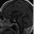
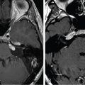
The patient is a 78-year-old male who was incidentally found to have a left jugular foramen mass consistent with meningioma. A 2-year observational period demonstrated persistent growth ( Figure 10.48.1 ), which led to consideration of treatment options. Prior to intervention, the patient had no cranial nerve deficits and had mild symmetric sensorineural hearing loss. In the setting of a growing tumor, normal lower cranial nerve function, older age, and significant cardiac comorbidities, the patient ultimately chose to undergo stereotactic radiosurgery (SRS) ( Figure 10.48.2 ).
| Radiosurgery Machine | Gamma Knife |
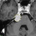
| Radiosurgery Dose (Gy) | 15.5 at the 50% isodose line |
| Number of Fractions | 1 |

A. Initial MRI 2 years prior to stereotactic radiosurgery (SRS): Axial T1-weighted image with gadolinium showing a homogenously enhancing jugular foramen mass with dural tails. B. Subsequent MRI 1 month prior to SRS: Axial T1-weighted image with gadolinium showing increased superior extent involving the proximal internal auditory canal. C. Subsequent MRI 1 month prior to SRS: Coronal T1-weighted image with gadolinium showing superior extention.

Imaging of the treatment plan. Yellow line, 16 Gy; green line, 8 Gy; pink line, cochlea outline.
| Critical Structure | Dose Tolerance |
|---|---|
| Brainstem | 15 Gy maximum point dose |
| Internal carotid artery in canal | Unknown dose tolerance |
| Cochlea | 4 Gy maximum point dose (extrapolated from vestibular schwannoma literature) |
| Lower cranial nerves in foramen | Unknown dose tolerance |
Related posts:
 Esthesioneuroblastoma – delayed postoperative radiosurgery for recurrence at long-term
 Null cell – delayed postoperative radiosurgery for growing perioptic residual
 Suprasellar non-small cell lung carcinoma metastasis – upfront radiosurgery
 Chondrosarcoma – definitive radiosurgery after subtotal resections
 Large vestibular schwannoma – delayed postoperative radiosurgery for growing residual
 Trigeminal neuralgia due to petroclival meningioma – upfront radiosurgery
Stay updated, free articles. Join our Telegram channel

Full access? Get Clinical Tree


