Jugular foramen/sigmoid sinus meningioma – postoperative radiosurgery for residual tumor
SKULL BASE REGION
Jugular foramen
HISTOPATHOLOGY
Meningioma, meningothelial type with bony invasion
PRIOR SURGICAL RESECTION
Yes
PERTINENT LABORATORY FINDINGS
None
Case description
The patient presented with a recurrent meningioma of the posterior fossa after prior resection via a retrosigmoid approach. Examination revealed moderate to severe ipsilateral sensorineural hearing loss, V2 and V3 distribution hypoesthesia, and normal lower cranial nerve function. Imaging revealed a locally aggressive mass consistent with meningioma, with significant osseous involvement ( Figure 10.47.1 ). The patient was offered surgical resection with adjuvant stereotactic radiosurgery (SRS) or external beam radiation. The patient ultimately underwent aggressive subtotal resection with posterior petrosectomy, resection of the thrombosed sigmoid sinus, and preservation of the lower cranial nerves ( Figure 10.47.2 ). Adjuvant SRS was performed months later ( Figure 10.47.3 ).
Radiosurgery Machine
Gamma Knife
Radiosurgery Dose (Gy)
16, at the 50% isodose line
Number of Fractions
1
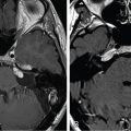
Figure 10.47.1.
A. Initial preoperative MRI: Axial T1-weighted image with gadolinium showing tumor involvement of the jugular foramen, pre- and post-sigmoid sinus dura. B. Initial preoperative CT: Axial cut without contrast showing significant hyperostosis and bony involvement.
Figure 10.47.2.
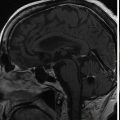
Postoperative MRI, prior to stereotactic radiosurgery: Axial T1-weighted image with gadolinium showing residual enhancement within the jugular foramen and along some remnant posterior fossa dura.
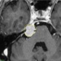
Figure 10.47.3.
Imaging of the treatment plan. Yellow line, 16 Gy; Green line, 8 Gy.
Critical Structure
Dose Tolerance
Brainstem
15 Gy maximum point dose
Internal carotid artery in canal
Unknown dose tolerance
Cochlea
4 Gy maximum point dose (extrapolated from vestibular schwannoma literature)
Lower cranial nerves in foramen
Unknown dose tolerance
Only gold members can continue reading. Log In or Register to continue
Apr 6, 2024 | Posted by drzezo in GENERAL RADIOLOGY | Comments Off on Jugular foramen/sigmoid sinus meningioma – postoperative radiosurgery for residual tumor


 Esthesioneuroblastoma – delayed postoperative radiosurgery for recurrence at long-term
 Null cell – delayed postoperative radiosurgery for growing perioptic residual
 Suprasellar non-small cell lung carcinoma metastasis – upfront radiosurgery
 Chondrosarcoma – definitive radiosurgery after subtotal resections
 Large vestibular schwannoma – delayed postoperative radiosurgery for growing residual
 Trigeminal neuralgia due to petroclival meningioma – upfront radiosurgery


