Urinary tract, a-p X-ray, i.v. urography
15 min after intravenous contrast
- 1: 12th rib
- Upper pole of right kidney
- Pelvis of right kidney
- Lower pole of right kidney
- Right ureter
- Renal papillae
- Fornix of minor calyx
- Minor calices
- Major calices
- Pelvis of left kidney
- Psoas major (lateral contour)
- Left ureter
- Urinary bladder
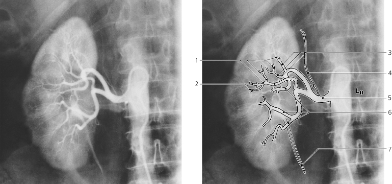
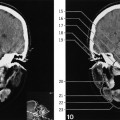
Renal artery, a-p X-ray, arteriography
- Arcuate arteries
- Interlobular arteries
- Interlobar arteries
- Inferior suprarenal artery
- Right renal artery
- Segmental arteries
- Right ureter
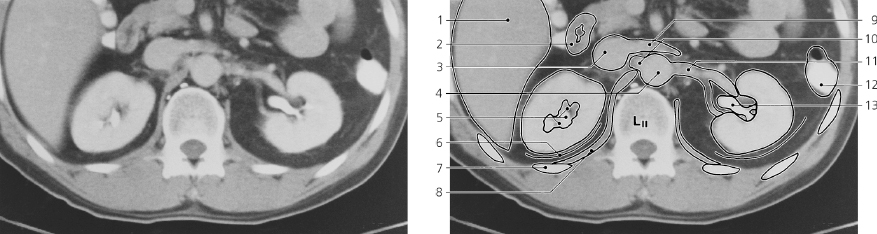
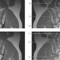
Kidneys, axial CT, after intravenous and peroral contrast
Only gold members can continue reading.
Log In or
Register to continue

Stay updated, free articles. Join our Telegram channel