• There is usually bilateral symmetrical involvement – unilateral involvement can occur with ascending infection or renal vein thrombosis • Papillae: the papilla initially swells and may eventually slough into the pelvicalyceal system causing colic, haematuria or hydronephrosis (as it appears as a filling defect it can mimic tumour, calculus or a blood clot) • Remaining defect: calyceal clubbing resulting from a flattened or concave pyramidal tip • Medullary nephrocalcinosis: this is usually the product of a metabolic disorder resulting in a raised serum calcium or a tubular defect resulting in hypercalciuria • Cortical nephrocalcinosis: this is seen in acute cortical necrosis of any cause • Bacteriuria, pyrexia and flank pain due to an ascending infection in 85% (usually E. coli), or haematogenous seeding in 15% (S. aureus) • The kidneys are the 2nd commonest site of TB involvement after the lung (even though the CXR is normal in 35–50% of cases it usually follows haematogenous spread from the lung) Although both kidneys are seeded, clinical manifestations are usually unilateral (> 70% of cases) • Early: an enlarged kidney – ‘Tuberculous autonephrectomy’: this may progress to homogeneous calcification within a dilated pelvicalyceal system so that the kidney appears as a lobulated calcified mass – Hydrocalycosis: a local calyceal dilatation due to a partial stricture of a major infundibulum (the infundibulum appears ‘cut off’ with a complete stricture) – Ureter: a ‘corkscrew’ appearance (due to multiple stenoses) – Early: trabeculation – Late: a thick-walled small-capacity bladder demonstrating calcification • Chronic renal parenchymal inflammation with replacement of the normal renal parenchyma with lipid-laden histiocytes • This is due to a fluke infestation (Schistosoma haematobium) • This is the most common cause of a non-atheromatous RAS and is the 2nd most common cause of an RAS (15–20%) • Medial fibroplasia is the commonest form, causing multiple short stenoses (with a ‘string of beads’ appearance on angiography) • It involves the distal main renal artery (and its major branches) Formally the gold standard • An aortogram is essential to demonstrate the number and location of the renal arteries and also the presence of any aortic or proximal renal vascular abnormalities • Selective arteriography should not be performed in RAS (except as a prelude to renal angioplasty) as it may cause renal artery dissection or occlusion • The kidney must be salvageable (e.g. a renal length > 8cm and a satisfactory GFR) • Indications: resistant hypertension • Complications (with a greater potential than seen in peripheral vascular disease): renal artery rupture and perforation • Renal vein occlusion by thrombus can be caused by: membranous glomerulonephritis (the commonest adult cause) • Acute: an enlarged kidney • Chronic: a normal or atrophic kidney • Thromboembolic occlusion of a renal artery usually leads to a focal renal infarction (less commonly total infarction)
Kidneys
RENAL PARENCHYMAL DISEASE
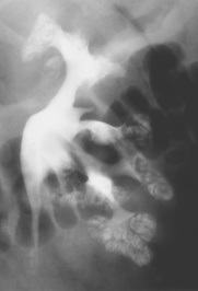
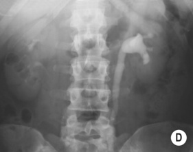
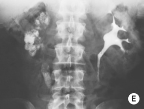
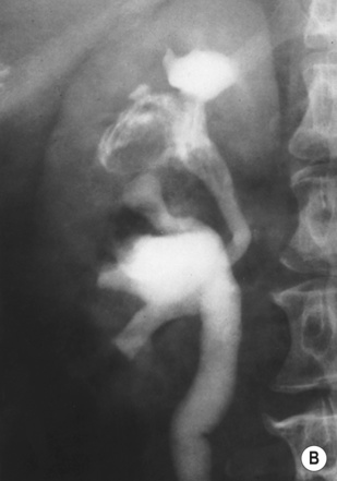
PAPILLARY NECROSIS
CT/IVU
 the papillary and calyceal changes are rarely uniform
 ‘Lobster claw deformity’: initially the necrotic areas erode the papilla tip and excavate from the fornices into the pyramids
 ‘Egg in cup’ appearance: with progression contrast curves around the papilla from both fornices
 if the papillae fail to separate (necrosis in situ) the calyces may appear normal
  the papillae may calcify (with spotty calcification in a ring or triangle around a translucent centre)
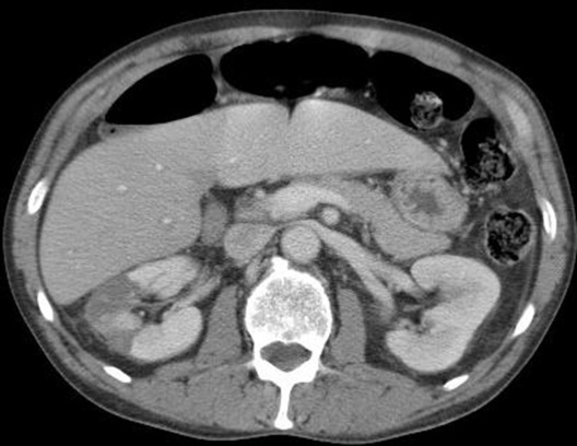
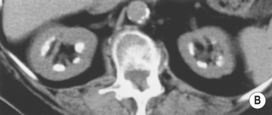
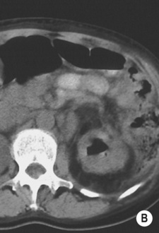
NEPHROCALCINOSIS
CT/IVU
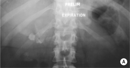
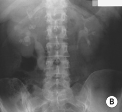
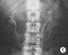
 calcification is usually bilateral and diffuse
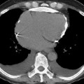
 calcification is usually punctate and patchy (classically with a ‘tramline’ appearance)
Glomerulonephritis  acute tubular necrosis
  acute cortical necrosis
No papillary or calyceal abnormality  no focal cortical loss
Papillary necrosis  medullary sponge kidney
  megacalycosis/polycalycosis
Papillary or calyceal abnormality  no focal cortical loss
Obstructive nephropathy  focal reflux nephropathy
Papillary or calyceal abnormality  focal cortical loss
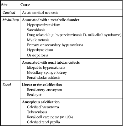
Site
Cause
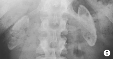
Cortical
Acute cortical necrosis
Medullary
Associated with a metabolic disorder
Hyperparathyroidism
Sarcoidosis
Drug related (e.g. hypervitaminosis D, milk-alkali syndrome)
Myelomatosis
Primary or secondary hyperoxaluria
Hyperhyroidism
Osteoporosis
Associated with renal tubular defects
Idiopathic hypercalciuria
Medullary sponge kidney
Renal tubular acidosis
Focal
Linear or rim calcification
Renal artery aneurysm
Real cyst
Amorphous calcification
Calcified haematoma
Tuberculosis
Renal cell carcinoma (in 10%)
Calcified renal papilla
RENAL TRACT INFECTION/INFLAMMATION
ACUTE PYELONEPHRITIS
DEFINITION
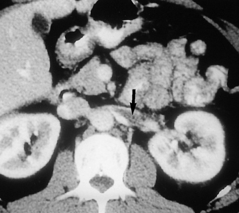
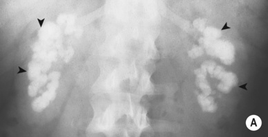
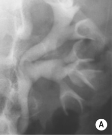
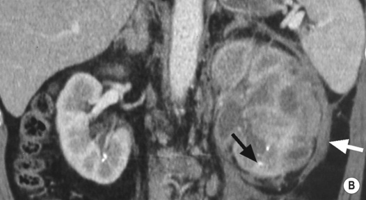
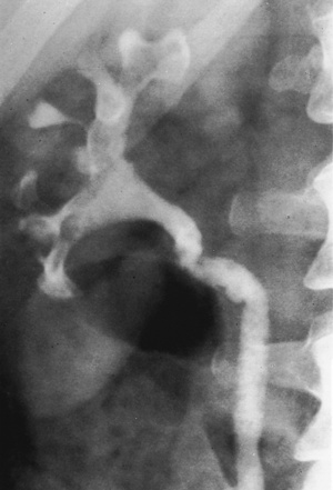
RENAL TUBERCULOSIS (TB)
DEFINITION
RADIOLOGICAL FEATURES
IVU/CT
 irregularity and destruction of ≥ 1 papillae (resembling papillary necrosis)
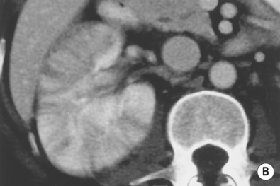
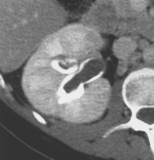
 Renal calcification (30%): this appears as punctate or curvilinear renal parenchymal calcification or as calcification within a caseous pyonephrosis (with a characteristic cloudy appearance in the distribution of the dilated calyces)
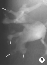
 Fibrotic strictures: these can occur anywhere within the renal tract
 a ‘pipestem’ appearance (due to a rigid and aperistaltic ureter)
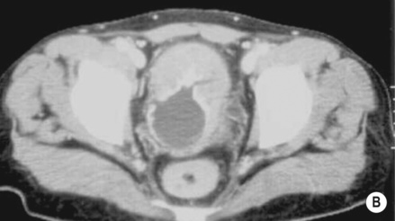
 bladder wall irregularity
  a slight decrease in capacity
 bladder TB is almost always associated with renal TB
  often there is VUR into a widely dilated upper tract
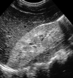
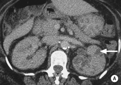
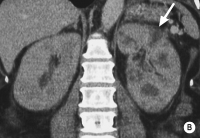
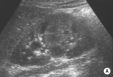
XANTHOGRANULOMATOUS PYELONEPHRITIS (XGP)
DEFINITION
 it is secondary to chronic urinary infection (e.g. E coli or Proteus mirabilis) and obstructing calculus disease
  it is associated with diabetes
SQUAMOUS METAPLASIA, LEUKOPLAKIA, MALACOPLAKIA, AND CHOLESTEATOMA
DEFINITION
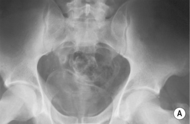
BILHARZIA (URINARY SCHISTOSOMIASIS)
DEFINITION
 the worm enters the skin and matures within the liver (via the portal vein)
  ova are then deposited within the bladder or ureteric submucosa (via the perivesical plexus)
  the ova produce an inflammatory reaction, leading to granuloma and stricture formation
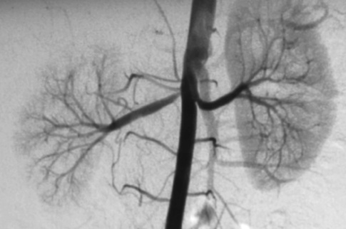
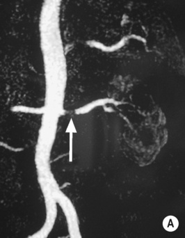
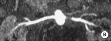
RENAL ARTERY STENOSIS (RAS)
RENAL ARTERY STENOSIS (RAS)
DEFINITION
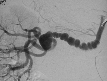
Fibromuscular dysplasia (FMD)
 it typically occurs in young women
 it can also affect the external iliac and carotid arteries
RADIOLOGICAL FEATURES
DSA
 it is increasingly being replaced by non-invasive methods
PEARLS
Renal angioplasty and stenting
 the best results are obtained with FMD
 renal failure
  flash pulmonary oedema
 branch occlusion
  occlusion of the main renal artery
  cholesterol emboli
  a short-term deterioration in renal function (due to the contrast medium given)
RENAL VASCULAR ABNORMALITIES
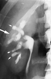
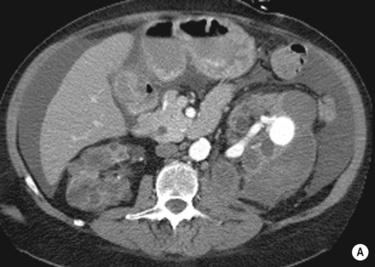
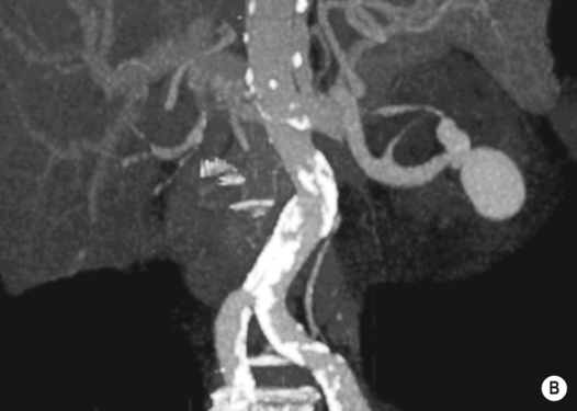
RENAL ARTERY ANEURYSMS
DEFINITION
RENAL VEIN THROMBOSIS
DEFINITION
 nephrotic syndrome
  dehydration
  hypercoagulable states
  renal or left adrenal tumours
RADIOLOGICAL FEATURES
IVU
 a faint or absent nephrogram
  absent pelvicalyceal filling (which may also be stretched and compressed by an oedematous renal parenchyma)
  rarely there may be an increasingly dense nephrogram (± striations)
 retroperitoneal venous collaterals can indent the PC system
RENAL INFARCTION
DEFINITION
 over time the infarcted area decreases in size with scar formation and tissue retraction
 it is usually primarily renal but may be part of a systemic vasculitis (e.g. SLE, PAN, Goodpasture’s or Wegener’s)
 no papillary or calyceal abnormality
 smooth, normal pelvicalyceal systems
  prominent renal sinus fat
 it usually follows an episode of severe ischaemia associated with hypotension, dehydration, or nephrotoxin exposure
 increased echogenicity within the cortex and pyramids
 little or no filling of the pelvicalyceal system
 the insult is more severe than that seen with ATN and is usually due to obstetric shock
 chronic: cortical calcification
 diabetes
  sickle-cell disease or trait
  obstruction complicated by infection
  acute infection
  haemophilia
  renal vein thrombosis
  acute renal failure in infancy

 it is usually bilateral (but can be unilateral or segmental)
 phaeochromocytoma
  horseshoe kidney
  Caroli’s disease
  hemihypertrophy
 cortical scarring (representing parenchymal loss) with underlying clubbed calyces
  localized scars are more common within the upper pole (R>L)
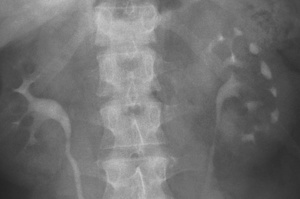

 vesicoureteric reflux (VUR)
  urinary obstruction
  pregnancy
  instrumentation
  immune deficiency
 it can however be useful if the diagnosis is in doubt or to exclude obstruction or abscess development
 delayed and poor pelvicalyceal system filling
  a dense, persistent or striated nephrogram (with severe disease)
 the affected segments are hypoechoic (they can be hyperechoic with haemorrhage)
  reduced corticomedullary differentiation (due to oedema)
  focal or diffuse reduced perfusion
 renal swelling with acute disease
 any abnormal parenchymal enhancement may persist for > 2 months and may develop into a scar
 emphysematous pyelonephritis
  xanthogranulomatous pyelonephritis
  cortical atrophy and renal failure
 fungal infections
  glomerulonephritis
  interstitial nephritis
 papillary ulceration occurs early, with later spread to the collecting system leading to fibrosis and stricture formation
 haematuria
  sterile pyuria
 calcification of the bladder, vas deferens and seminal vesicles is rarely seen
 widespread cavitations may mimic hydronephrosis (but the pelvis and infundibula of the calyces are not dilated unless there is an associated obstruction)
 it is associated with diabetes and obstruction
 there is a high mortality rate (> 60%) and it may require nephrectomy
 it has a lower mortality rate, and percutaneous drainage and antibiotics may be sufficient
 the patient is usually diabetic
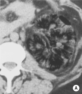

 the focal form can mimic a tumour
 an enlarged (global or focal) non-excreting kidney
  dilated affected calyces with internal echoes or debris
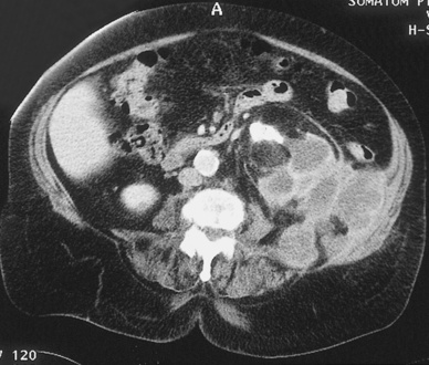
  a heterogeneous kidney with multiple non- or rim-enhancing low attenuation areas (−15 to −20 HU) representing dilated calyces and xanthomas
  perinephric extension (± a thickened Gerota’s fascia)
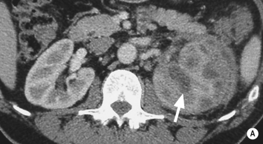
 there are enhancing thick irregular walls (± perinephric inflammatory change)
  gas within the lesion is diagnostic
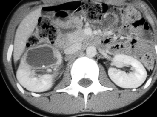
 absent renal function
 this may extend in any direction
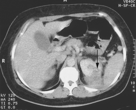
 it is associated with recurrent urinary tract infections and calculi
 it appears as thickened folds and irregularity of the renal pelvis
 small plaques are visible on the mucosal surfaces (bladder > upper urinary tract)
 there may be impaired renal function if the parenchyma is involved
 it is associated with urinary tract infection and chronic obstruction
 there can be multiple filling defects (representing granulomas or ureteritis cystica)
  parallel lines of ureteric calcification can be seen
 diabetics are at increased risk
 it is highly echogenic on US, and it may demonstrate air within the collecting system and bladder
 there is a risk of septicaemia and endotoxic shock (especially with attempted drainage)
 debris or gas (dense shadowing) within the renal pelvis
  cortical loss or a perinephric abscess if long-standing
 there may be a renal or perinephric abscess
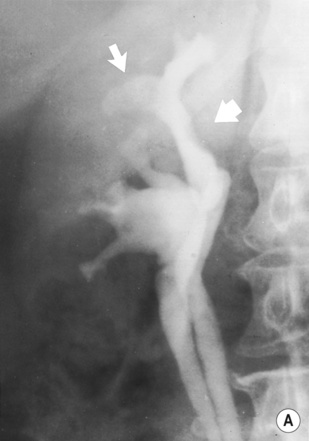

 the resultant increased renin (and angiotensin II) levels leads to vasoconstriction
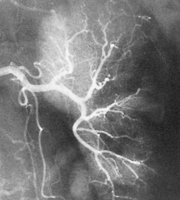
 atheroma involves the origin (ostial) or proximal ⅓ of the renal artery
  there can be post-stenotic dilatation
 renal arterial lesions are caused by eccentric atheromatous plaques of the proximal renal artery
 aortic disease may consist of diffuse or focal stenoses, occlusion, or a fusiform AAA
 a renal artery to aortic velocity ratio (RAR) > 3.5
  a ‘parvus and tardus’ effect within the intrarenal vessels (due to velocity reduction and slowing of the acceleration of the systolic upstroke)
 it has a sensitivity (97%) and specificity (92%) comparable with intra-arterial angiography for detecting stenoses within the main and segmental arteries


 it may be associated with Williams’ syndrome or neurofibromatosis
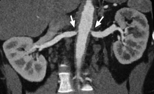
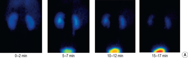
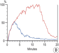
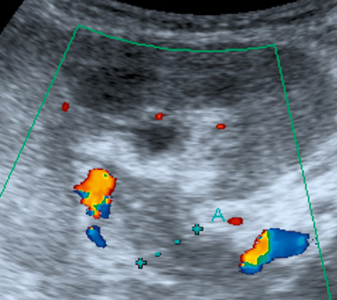
 blue line, left kidney).*
 mycotic
  post-traumatic
  atherosclerotic
  vasculitic
  fibromuscular hyperplasia
 both will demonstrate aneurysmal dilatation
 absent wall calcification
  occurring during pregnancy
 aneurysms tend to be more peripheral than with FMD
 it may lead to impaired renal function due to a ‘steal’ effect
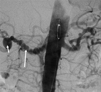
 with chronicity venous collaterals can open with only slightly impaired renal function
 renal vein thrombus and a lack of flow within the main veins (± reversed end diastolic flow within the parenchymal veins)
 prolonged irregular parenchymal enhancement
  prolonged corticomedullary differentiation
  the ‘cortical rim’ sign
  delayed or absent contrast medium excretion
 3D gadolinium-enhanced MRA may differentiate benign from tumour thrombus
 selective renal venography may identify any filling defects
 aortic aneurysm
  trauma
  thrombosis (due to atheroma or vasculitis)
 this can progress to a shrunken end-stage kidney
 there is failure of tracer uptake on later images
 T1WI + Gad: no enhancement