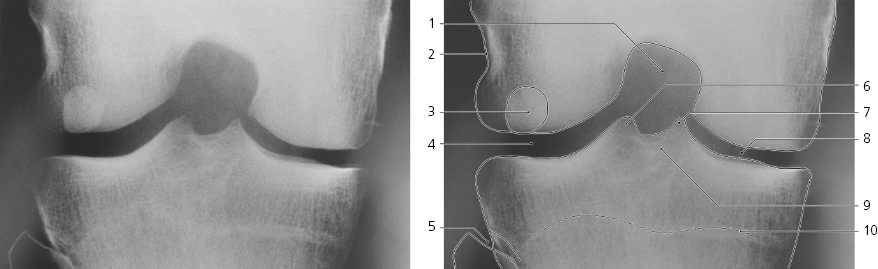
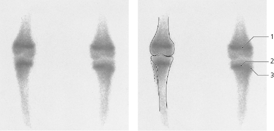
Knee, 99m Tc-MDP, a-p scintigraphy, child 12 years
Growth plate of distal epiphysis of femur
Growth plate of proximal epiphysis of tibia
Growth plate of proximal epiphysis of fibula
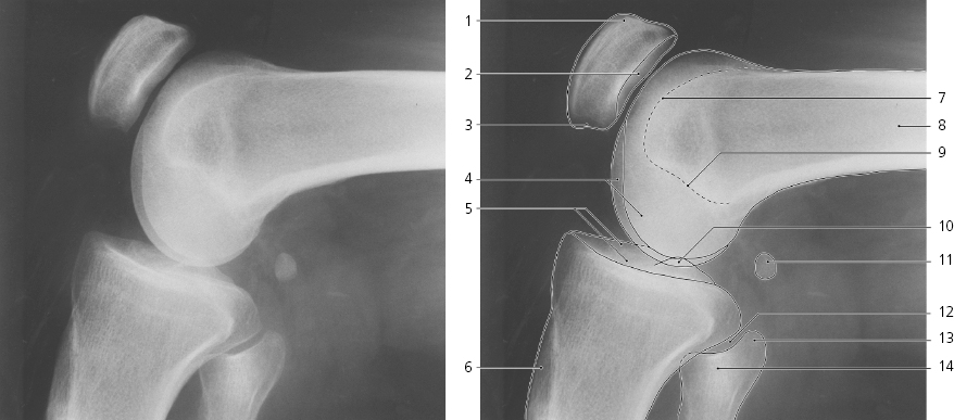
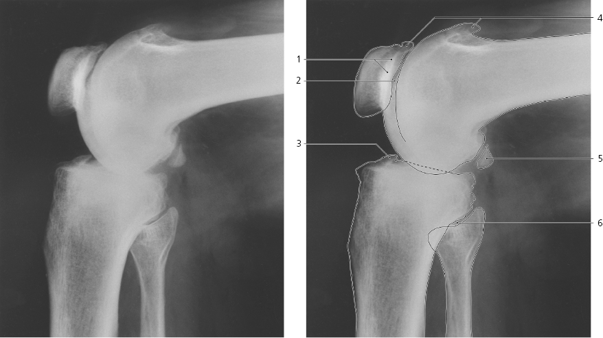
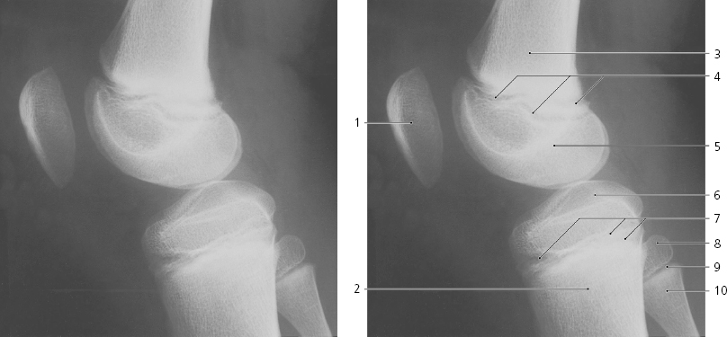
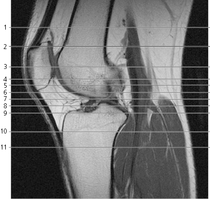
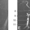
Scout view of knee
Lines #1–11 indicate position of sections in the following axial MR series. Interpretation of the scout image can be found in the sagittal series, page 132, image # 8.
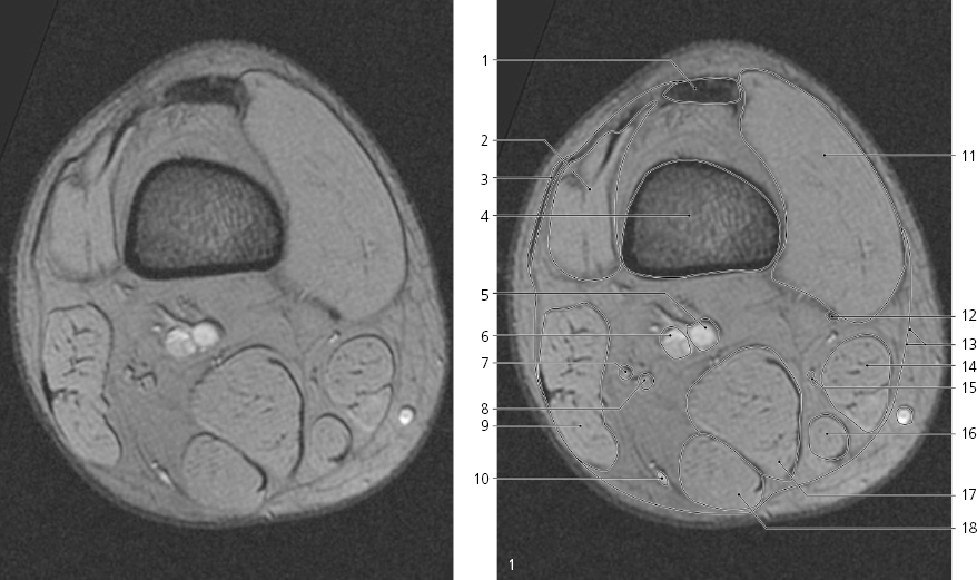
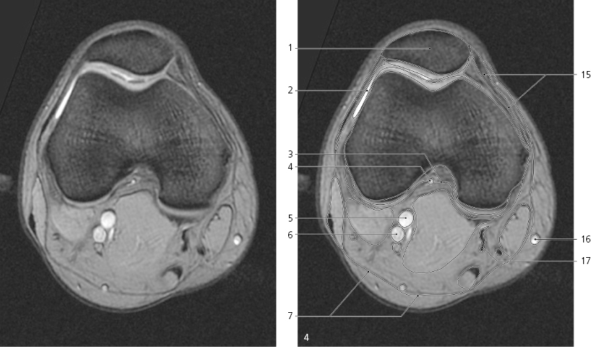
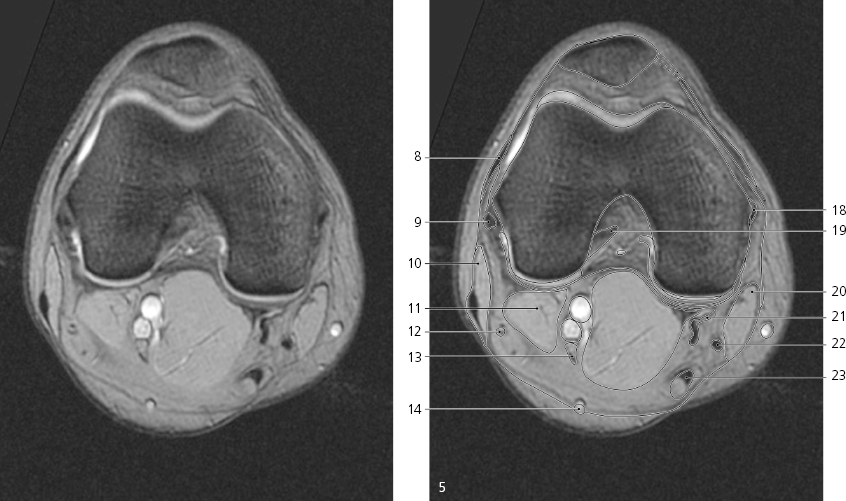
Knee, axial MR
Quadriceps tendon
Vastus lateralis →
Iliotibial tract →
Femur (shaft)
Popliteal artery →
Popliteal vein →
Common peroneal nerve →
Tibial nerve →
Biceps femoris →
Small saphenous vein →
Vastus medialis →
Adductor magnus (tendon) →
Fascia lata →
Sartorius →
Saphenous nerve →
Gracilis →
Semimembranosus →
Semitendinosus →
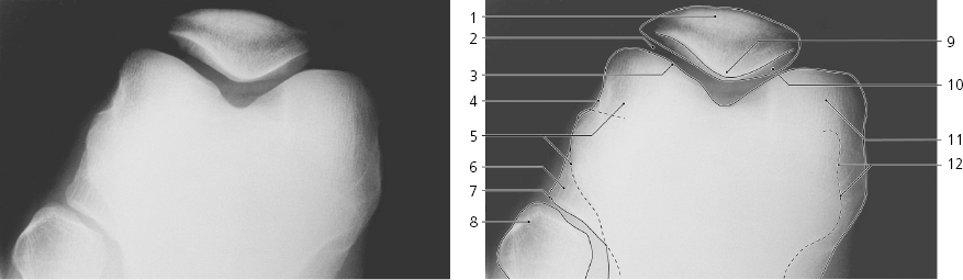
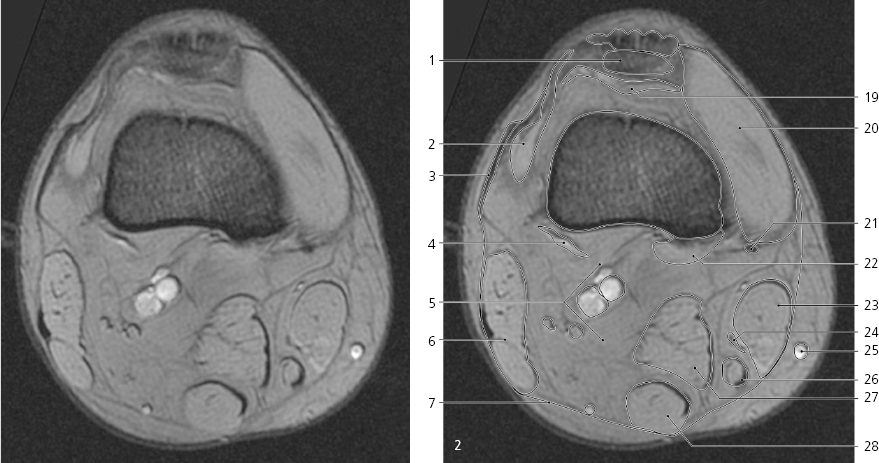
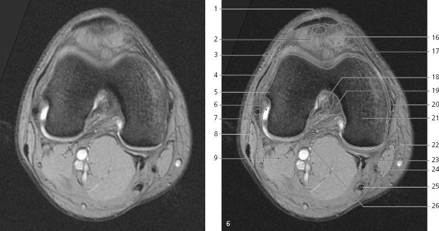
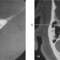
Knee, axial MR
Scout view on page 122
Patella (basis) →
Vastus lateralis ↔
Iliotibial tract ↔
Plantaris →
Popliteal fossa →
Biceps femoris →
Popliteal fascia →
Patella →
Patellofemoral joint cavity (with synovia)
Lateral retinaculum patellae →
Articular cartilage of patella and femur
Iliotibial tract ↔
Plantaris muscle →
Biceps femoris ↔
Lymph node
Common peroneal nerve ↔
Tibial nerve ↔
Small saphenous vein ↔
Suprapatellar bursa
Vastus medialis ←
Adductor magnus (tendon) ←
Gastrocnemius (medial head) →
Sartorius ↔
Saphenous nerve ←
Great saphenous vein ↔
Gracilis ↔
Semimembranosus ↔
Semitendinosus ↔
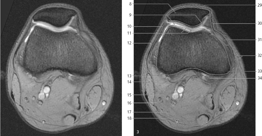
Quadriceps femoris (tendon)
Medial patellofemoral ligament
Vastus medialis ←
Fascia lata ↔
Adductor tubercle (adductor magnus insertion)
Bursa
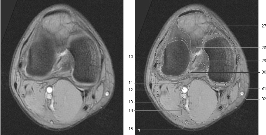
Knee, axial MR
Scout view on page 122
Patella ↔
Synovia in joint cavity
Posterior joint capsule
Median articular artery
Popliteal artery ↔
Popliteal vein ↔
Popliteal fascia ↔
Iliotibial tract ↔
Fibular (lateral) collateral ligament →
Biceps femoris ↔
Plantaris and gastrocnemius (lateral head)
Common peroneal nerve ←
Tibial nerve ↔
Small saphenous vein ↔
Medial patellofemoral ligament ↔
Great saphenous vein ↔
Semimembranosus (tendon) ↔
Tibial (medial) collateral ligament →
Anterior cruciate ligament →
Sartorius ↔
Semimembranosus (tendon) ↔
Gracilis ↔
Semitendinosus ↔
Knee, axial MR
Only gold members can continue reading. Log In or Register to continue