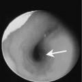
Chapter 107 A laryngocele is an abnormal cystic dilatation of the laryngeal ventricle with a persistent the communication between the cyst and the laryngeal ventricle. These lesions may be filled with air or fluid. Laryngoceles are classified as internal, external, or mixed. Internal laryngoceles are confined to the endolarynx, whereas an external laryngocele extends through the thyrohyoid membrane into the soft tissues of the neck. This extension usually occurs through the natural opening in the thyrohyoid membrane for the superior laryngeal artery. A laryngocele with both internal and external components is termed a mixed laryngocele. Laryngoceles may be congenital or acquired and are seen in all ages. In children, laryngoceles are thought to be congenital and are a cause of stridor in newborn and young children. Laryngoceles may be unilateral or bilateral. There is no reported gender predilection. In adults, laryngoceles may be idiopathic in origin. In the past they have been associated in adults who are trumpet players or glass blowers. However, in our experience, the most common association in adults is squamous cell carcinoma involving the true vocal cords. Two to six percent of glottic squamous cell carcinomas are associated with laryngocele formation. The clinical findings often depend on the size and extent of the lesion. Children may present with signs of respiratory distress such as inspiratory stridor, intercostal retractions, and episodes of cyanosis. Patients may have intermittent hoarseness or aphonia or a muffled cry. Associated anomalies include laryngomalacia and vocal cord paresis. At endoscopy, laryngoceles appear as soft rounded submucosal soft tissue masses that may involve the pyriform sinus, aryepiglottic fold, pyriform sinus, glottis, and hypopharynx (Fig. 107–1). These lesions are mucosally covered and have a characteristic bluish hue. Large lesions may extend into and obstruct the laryngeal lumen. The diagnosis of laryngocele is confirmed if the lesion is seen to inflate and deflate at endoscopy.
Laryngocele
Epidemiology
Clinical Findings
Related posts:


Stay updated, free articles. Join our Telegram channel

Full access? Get Clinical Tree


