Fig. 13.1
View of the larynx as provided by the 70 ° rigid scope
Before the procedure, local anesthesia with tetracaine (Pontocaine) 2% or lidocaine 4% with addition of Adrenalin is applied. If the transnasal route is used, a nasal decongestant with local anesthetic (e.g., xylometazoline with lidocaine) is highly recommended, if the transoral route is used, oral cavity, pharynx, and laryngeal mucosa are sprayed step by step; the larynx is then brushed with an arcuated cotton-bearing hook under indirect vision. With extreme gagging, infiltration anesthesia of the cranial recurrent nerve may be performed from outside, and infiltration of the glossopharyngeal nerve at the posterior inferior pole of the tonsil with 2 cc of 1% lidocaine may be necessary. Cocaine is effective in cases of inadequate action of other local anesthetics and is tolerated well by the laryngeal mucosa. The patient is usually sitting upright. Special instruments, such as an arcuated cotton-bearing hook, epiglottic hook (Reinhardt), cutting forceps, binocular microscope, or laryngoscope, may be used.
Visualization should follow a standardized pattern as to guarantee inspection of all regions of the larynx. Inspection starts at the tongue base bilaterally, then the valleculae and epiglottis are inspected. The aryepiglottic folds are followed posteriorly, then the arytenoid cartilages are inspected. Attention is now directed toward the glottis: The false vocal cords, the sinus, and the true vocal cords are inspected during respiration and phonation. During respiration, the subglottic space can be visualized. Indirect laryngoscopy offers only limited view of the piriform sinus and the postcricoid region. These regions can only be properly visualized by direct laryngoscopy under general anesthesia.
Follow-up after surgery may include voice rest for 3–7 days and abstinence from smoking and alcohol. First food ingestion may occur 2 h after surgery. Inhalation (e.g., dexpanthenol) may ease a feeling of dryness and sore throat. If needed, antibiotic coverage and medical suppression of cough for 3 days may be used. If the patient is prone to edema or has a narrow glottic opening, monitoring and corticosteroids may be necessary.
Risks and complications include hoarseness (due to tissue defects, vocal cord trauma, scarring of the vocal cords, or formation of synechias) and laryngeal edema with dyspnea.
Patient and surgeon should be relaxed and calm. Scopes and instruments may be handled more easily, if the surgeon’s upper arms are supported by his thorax. A tremorous hand as well as a nervous patient may lead to defective excision and damage of the larynx. Before the excision, the patient should take a couple of deep breaths in order to allow the larynx to stay still during prolonged inspiration.
Advantages of the indirect laryngoscopy comprise the cost-effectiveness with no need of general anesthesia, no need of assistance and outpatient procedure, and no need of costly equipment. The disadvantages are the limited indications and practicality, the limitation to the use of one hand, lack of precision, and dependence on the patient’s cooperation.
Direct Rigid Laryngoscopy
Direct vision (direct laryngoscopy) offers the advantage of three-dimensional appreciation of the larynx, bimanual instrumentation, application of tools such as the laser, and clear visualization of “hidden” regions such as the piriform sinus and the postcricoid region. Due to the usage of a rigid scope, general anesthesia is mandatory. Rigid laryngoscopy is mostly part of a so-called panendoscopy including a flexible or rigid bronchoscopy and a flexible or rigid esophagoscopy.
Preoperative management includes proper documentation of the pathology in question and the status quo of laryngeal anatomy and physiology, preferably by videostroboscopy (Table 13.1). Management by anesthesia requires atraumatic intubation with a small endotracheal tube, typically a 5.0 tube. Most cases can be managed with the endotracheal tube in place. If necessary, jet ventilation can be used. Directly at the end of surgery, application of 4% lidocaine is prudent to minimize the risk of laryngospasm after extubation.
Table 13.1
Indications and contraindications for direct/indirect laryngoscopy
Indications for direct laryngoscopy |
Persistent hoarseness |
Suspected neoplasm |
Chronic cough |
Chronic postnasal drainage |
Recurrent epistaxis |
Chronic rhinorrhea |
Chronic nasal congestion or obstruction |
Hemoptysis |
Hemorrhage from throat |
Throat pain |
Otalgia |
Airway obstruction |
Dyspnea |
Stridor |
Dysphagia |
Head or neck masses – unknown primary tumor |
Laryngeal injury with hoarseness or airway obstruction |
Chronic aspiration |
Velopharyngeal incompetence |
Suspected foreign body |
Recurrent serous otitis media in an adult |
History of tobacco use |
Obstructive sleep apnea or severe snoring |
History of thyroid disorders |
Anosmia/Hyposmia |
Headaches or facial pain |
Relative contraindications for direct laryngoscopy |
Problems with stiffness of cervical spine |
Retrognathia |
Trismus |
Different rigid laryngoscopes are available (Kleinsasser laryngoscope, Boston, Dedo, Ossoff, Zeitels, etc.). Proper positioning of the patient is the first step to good visualization during the procedure. The patient is in supine position, and the neck is extended using a pad under the shoulders and a soft ring under the head to attain stable positioning (Fig. 13.2).
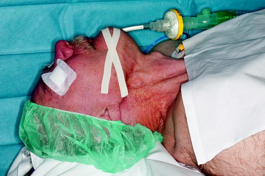
Fig. 13.2
Rigid laryngoscope (Kleinsasser) with included lighting
Exposure is key to visualization and proper performance of surgical procedures. Different means of instrumentation (rigid laryngoscopes) are available, all offering excellent exposure Peroral rigid laryngoscopy begins with application of a tooth guard (Fig. 13.3), preferably using soft plastic dental protection with a metal coverage to prevent accidental damage to the teeth. The upper incisors are at greatest risk of injury as they are long and exposed to the rigid instrument. It is extremely important never to use the teeth as a fulcrum. Care is also taken not to injure the lips by pulling them inside with the laryngoscope and lacerating them between the scope and the teeth. Gentle insertion of the laryngoscope with lighting is performed with the left hand in the right-handed surgeon (Fig. 13.4). The right hand is then free for the usage of suction or a grasping instrument to aid proper exposure. Tongue and soft palate including the tonsils are inspected. Next the valleculae, tongue base and the lingual and laryngeal surface of the epiglottis are visualized. The aryepiglottic folds are followed posteriorly to visualize the arytenoid cartilages. The piriform sinus is inspected bilaterally. The endotracheal tube is then pushed anteriorly, and the postcricoid area is inspected thoroughly. The laryngoscope is used to lift the tip of the epiglottis. This so-called “engagement” of the epiglottis is necessary to gain clear visualization and access to the larynx. Now the attention is turned toward the larynx and the microscope is used for the following part of the procedure (Fig. 13.5). Proper inspection is performed including the false and true vocal cords; the ventriculus (sinus of Morgagni) is palpated with a hook. The anterior and posterior commissures are inspected. If the anterior commissure cannot be visualized, gentle cricoid pressure can be applied to maneuver the larynx in the proper position. The laryngoscope is retracted slowly; the tooth guard is removed. Finally, tongue base, floor of the mouth, lateral pharyngeal walls, and the lower part of the nasopharynx are palpated enorally and bimanually, using one finger enorally and the opposite hand from the outside.
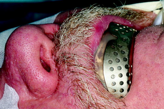
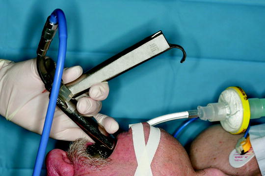
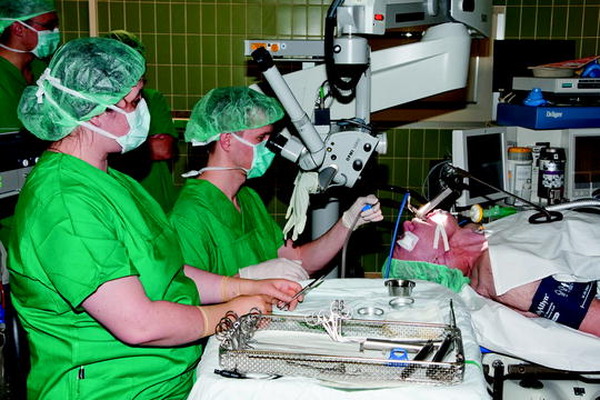
Fig. 13.3
Proper patient positioning
Fig. 13.4
Application of tooth guard and gentle insertion of the laryngoscope
Fig. 13.5
Laryngoscopy in position and view through the microscope
Diagnostic Indications for Direct Laryngoscopy
Related posts:


Stay updated, free articles. Join our Telegram channel

Full access? Get Clinical Tree


