Left-to-Right Cardiovascular Shunts


Left-to-Right Cardiovascular Shunts


Ramesh S. Iyer, MD

Mark R. Ferguson, MD


INTRODUCTION

There are several ways of categorizing congenital heart disease. One method that we use, described by Higgins, relies upon a few features that the radiologist should either elicit from the medical record or comment on in the imaging report1:


  • Is the patient cyanotic or acyanotic?


  • Is pulmonary vascularity increased or low/normal? The appearance of pulmonary arterial overcirculation, or “shunt vascularity,” was characterized in Chapter 9. Determining whether there is enlargement and abundance of pulmonary arteries takes experience. Identifying low pulmonary vascularity is perhaps even more challenging (Fig. 10.1). Fortunately for the radiologist, in the cyanotic child, both low and normal pulmonary arterial flow are managed similarly in that they both exhibit pulmonary-systemic (right-to-left) shunting. Therefore, the question for radiography interpretation boils down to whether or not pulmonary vascularity is increased (Fig. 9.6).1,2


  • Is there cardiomegaly or a normal heart size?

Using these parameters, most cardiac shunt pathologies can be divided into one of four categories (Table 10.1).1 The focus of this chapter will be group 1 lesions: left-to-right shunts. Cyanotic lesions (groups 2-4) are discussed in Chapter 11.

Left-to-right shunts are characterized by pulmonary arterial overcirculation and lack of cyanosis. When cardiomegaly is present, the amount of cardiac enlargement is generally proportional to the increase in pulmonary vascularity.1 Assessing left atrial enlargement is useful for distinguishing between left and right shunts. If the left atrium is enlarged, then the patient likely has a shunt that is distal to the level of the atria, most often a ventricular septal defect (VSD). If the left atrium is normal in size in the setting of shunt vascularity, then the child likely has an atrial septal defect (ASD) that allows for decompression of the left atrium (note, a VSD may also be present). On a frontal chest radiograph, signs of left atrial enlargement include widening of the carinal angle beyond 90 degrees and a “double-density” appearance of the right atrium because of the superimposed left atrium enlarging downward and rightward (Fig. 9.7A).3 On the lateral view, the left atrium forms the posterior cardiac margin. The enlarged left atrium displaces the left main bronchus posteriorly and projects
over the thoracic spine (Fig. 9.7B). On echocardiography, CT, or MR, the left-to-right shunt may be directly visualized, and quantification of flow across the defect may be performed with MR or echo by calculating the ratio of flow through the main pulmonary artery (Qp) versus flow through the ascending aorta (Qs). Another method for predicting the type of shunt present is the age at presentation. VSDs and atrioventricular septal defects (AVSDs) present early in life, usually in infancy. ASDs typically present in preschool-aged children or in older children, even presenting as late as in adulthood (“A” in ASD for Adults). Patent ductus arteriosus (PDA) usually presents in premature neonates and makes up the majority of left-to-right shunts in these patients.4


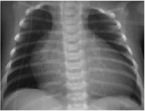
FIG. 10.1 • Pulmonary hypovascularity in a newborn with Ebstein anomaly. AP chest radiograph demonstrates decreased pulmonary vascularity and cardiomegaly. The prominent right heart margin represents right atrial enlargement.


Table 10.1 CLASSIFICATION OF SHUNT LESIONS


Group 1 lesions: Acyanotic; pulmonary arterial overcirculation (left-to-right shunts)


Atrial septal defect (ASD)


Partial anomalous pulmonary venous return (PAPVR)


Atrioventricular septal defect (AVSD), endocardial cushion defect (ECD)


Ventricular septal defect (VSD)


Patent ductus arteriosus (PDA)


Other aortic level shunts (e.g., aortopulmonary window, ruptured sinus of Valsalva aneurysm)


Group 2 lesions: Cyanotic; decreased pulmonary vascularity; no cardiomegaly (tetralogy of Fallot and its variants)


Tetralogy of Fallot


Transposition with pulmonic stenosis and VSD


Double-outlet right ventricle with pulmonic stenosis and VSD


Doublet-outlet left ventricle with pulmonic stenosis and VSD


Single ventricle (univentricular atrioventricular connection) with pulmonic stenosis


Corrected transposition with pulmonic stenosis and VSD


Pulmonic atresia with intact ventricular septum, type 1


Pulmonic stenosis with AVSD


Hypoplastic right ventricle syndrome


Some types of tricuspid atresia (large ASD and pulmonic stenosis or atresia)


Group 3 lesions: Cyanotic; decreased pulmonary vascularity; cardiomegaly (right heart lesions)


Ebstein anomaly


Pulmonary stenosis (critical) with ASD or patent foramen ovale (PFO)


Some types of tricuspid atresia (restrictive ASD)


Pulmonic atresia with intact ventricular septum, type 2


Transient tricuspid regurgitation of the newborn


Group 4 lesions: Cyanotic; pulmonary arterial overcirculation (admixture lesions, “T” lesions)


Transposition of great arteries


Truncus arteriosus


Total anomalous pulmonary venous return (TAPVR)


Tricuspid atresia


Single ventricle (univentricular atrioventricular connection)


Double-outlet right ventricle


Double-outlet left ventricle


Atrioventricular septal defect (complete form)


Hypoplastic left heart syndrome


Pulmonary atrioventricular fistula


Group 1 is separated from the remaining groups by including only acyanotic lesions. Groups 2 and 3 are distinguished by heart size—group 2 lesions have normal heart size, while group 3 pathologies exhibit cardiomegaly. Group 4 shunts feature both cyanosis and pulmonary arterial overcirculation, which are incongruous and indicate an admixture lesion.1


Adapted and reprinted with permission from: Higgins CB. Radiography of congenital heart disease. In: Webb WR, Higgins CB, eds. Thoracic Imaging: Pulmonary and Cardiovascular Radiology. 2nd ed. Philadelphia, PA: Lippincott Williams & Wilkins; 2011:742-767.


VENTRICULAR SEPTAL DEFECT

VSD is the most common cardiac anomaly identified in children, accounting for 20% to 40% of all cardiac malformations.5, 6, 7 and 8 The lesion may be an isolated finding, or a component of more complex cardiac disease such as tetralogy of Fallot or AVSD. There may also be associated vascular pathology such as transposition of great vessels or aortic coarctation.5 The precise incidence of VSD is difficult to ascertain given the relatively high spontaneous closure rate. Twenty five to forty percent of VSDs close spontaneously by the age of 2 years, and 90% of defects that will eventually close do so by the age of 10 years.6,9

There are four types of VSDs. The majority of VSDs are of the perimembranous subtype (70% to 80%). Perimembranous defects are located within the left ventricular outflow tract immediately below the aortic valve. The membranous septum is the site of fusion for multiple components during embryologic development of the ventricular septum, rendering this location most susceptible to malformation. Muscular defects (10%) may be single or multiple—the latter producing a “Swiss cheese” appearance—and are the result of excessive muscular resorption during fetal life. Inlet or AV canal defects (10%) involve the posterior and inferior ventricular septum. Finally, outlet or supracristal VSDs (5%) are situated high within the ventricular outflow tract at the joined annulus between aortic and pulmonic valves.1,3,5,6,10, 11, 12, 13 and 14

The pathophysiology of a VSD depends upon the size of the defect and pulmonary vascular resistance. During systole, blood within each ventricle may either exit the normal outflow tract or cross the VSD and exit the heart via the outflow tract of the other ventricle. If the VSD is moderate to large in size, then blood will travel from the high-pressure left ventricle across the VSD into the right ventricle and enter the low-resistance pulmonary arterial bed. However, over time, systemic pressures get transmitted to the pulmonary vascular system and the latter pressures increase. Eventually, pulmonary arterial resistance will equal or exceed systemic vascular resistance, and right-to-left shunting will occur. This shunt reversal physiology is referred to as Eisenmenger syndrome.1,6,7

On chest auscultation, VSDs produce a holosystolic murmur usually accompanied by a palpable thrill.5,6 Children with small defects are otherwise asymptomatic. Children with moderate to large defects typically present during the first few months of life. These patients are acyanotic and exhibit signs of congestive heart failure including tachypnea and tachycardia, and failure to thrive.5,10 By contrast, older children or adolescents with Eisenmenger syndrome have cyanosis and clubbing and usually no murmur.5


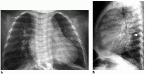
FIG. 10.2 • VSD in a 5-month-old boy. A: AP chest radiograph shows cardiomegaly and increased pulmonary arterial flow. B: Lateral projection illustrates left atrial enlargement, with the posterior cardiac margin projecting over the spine (arrow).

On chest radiography, moderate to large VSDs produce cardiomegaly and increased pulmonary arterial vascularity in infants (Fig. 10.2). The left atrium, right atrium, and right ventricle can all be enlarged. Small VSDs may yield a normal chest radiograph. Echocardiography is usually sufficient for imaging VSDs, offering both precise localization of the lesion and shunt quantification across the defect. Both MR and CT are useful modalities for VSD characterization if acoustic windows are poor (Fig. 10.3).3,4,7,10,12, 13 and 14

May 24, 2016 | Posted by in PEDIATRIC IMAGING | Comments Off on Left-to-Right Cardiovascular Shunts

Full access? Get Clinical Tree

Get Clinical Tree app for offline access