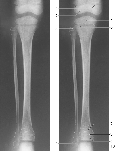
Leg, a-p X-ray
- Lateral condyle of femur
- Lateral condyle of tibia
- Apex of fibula
- Head of fibula
- Neck of fibula
- Shaft of fibula
- Nutrient canal
- Compact bone of tibial shaft
- Medullary cavity of tibia
- Fibular notch of tibia (syndesmosis)
- Lateral malleolus
- Medial condyle of femur
- Superior articular surface of tibia
- Medial condyle of tibia
- Medial and lateral tubercle
- Shaft of tibia
- Medial malleolus
- Trochlea of talus

Leg, child 6 years, a-p X-ray
- Growth plate
- Distal epiphysis of femur
- Proximal epiphysis of fibula
- Distal epiphysis of fibula
- Proximal epiphysis of tibia
- Growth plate
- Harris lines (signs of temporary growth arrest)
- Growth plate
- Distal epiphysis of tibia
- Talus
Only gold members can continue reading.
Log In or
Register to continue

Stay updated, free articles. Join our Telegram channel


