and Zdeněk Fryšák1
(1)
Department of Internal Medicine III – Nephrology, Rheumatology and Endocrinology, Faculty of Medicine and Dentistry, Palacky University Olomouc and University Hospital Olomouc, Olomouc, Czech Republic
Keywords
Hypoechoic solid noduleEstimated risk of malignancyFine-needle aspiration biopsy13.1 Essential Facts
Up to 55% of benign nodules are hypoechoic compared to thyroid parenchyma; hypoechogenicity alone, therefore, is not a diagnostic sign of malignancy. In addition, benign nodules ≤1 cm are more likely to be hypoechoic than larger nodules [1].
According to the 2015 American Thyroid Association (ATA) Guidelines [1]:
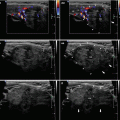
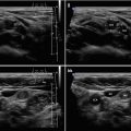
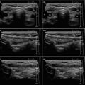
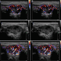
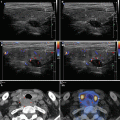
Intermediate suspicion of malignancy (estimated risk 10–20%) is attached to a hypoechoic solid nodule with a smooth regular margin (Fig. 13.1aa), but without microcalcifications, extrathyroidal extension, or “taller-than-wide” shape. This appearance has the highest sensitivity (60–80%) for papillary thyroid carcinoma (PTC), but a lower specificity than the preceding high suspicion pattern, and FNAB should be considered for these nodules ≥1 cm to refute malignancy.
Low suspicion malignancy risk (estimated risk 5–10%): isoechoic or hyperechoic solid nodule, or partially cystic nodule with eccentric uniformly solid areas without microcalcifications, irregular margin or extrathyroidal extension, or “taller-than-wide” shape prompts low suspicion for malignancy. Only about 15–20% of thyroid cancers are isoechoic or hyperechoic on US, and these are generally the follicular variant of PTC or follicular thyroid carcinoma (FTC). Fewer than 20% of these nodules are partially cystic. Therefore, these appearances are associated with a lower probability of malignancy and observation may be warranted until the size is ≥1.5 cm.


Fig. 13.1
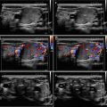
(aa) A 58-year-old women with a suspicious small solid nodule (arrowheads) in the RL, size 12 × 8 × 6 mm and volume 0.3 mL. Post-thyroidectomy histology: benign hyperplastic nodule. US overall view: elliptical shape; homogeneous; hypoechoic; without microcalcifications; well-defined margin; tiny nonsuspicious nodule in the LL—homogeneous, isoechoic with halo sign; Tvol 12 mL, RL 7 mL, and LL 5 mL; transverse. (bb) Detail of suspicious small solid nodule (arrowheads), CFDS: diffusely increased vascularity, pattern II; transverse. (cc) Detail of suspicious small solid nodule (arrowheads): homogeneous structure; hypoechoic; well-defined margin; longitudinal. (dd) Detail of suspicious small solid nodule (arrowheads), CFDS: increased peripheral vascularity and one intranodal vessel branch, pattern II; longitudinal
Stay updated, free articles. Join our Telegram channel

Full access? Get Clinical Tree


