12 Lesions with Left-to-Right Shunt
With Christian J. Kellenberger
Definition and Classification
 A spectrum of lesions with either an intracardiac or extracardiac communication whose fundamental abnormality is allowing blood to shunt from the higher pressure systemic circulation to the lower pressure pulmonic circulation, resulting in oxygenated blood recirculating through the lungs
 Levels of shunt
- • Intracardiac
- – Atrial septal defects (Fig. 12.1)
- ○ Patent foramen ovale: persistence of fetal pathway from the right atrium to the left atrium
- ○ Fossa ovalis defect (also called secundum defect): one or more holes in the floor of the fossa ovalis
- ○ Primum defect: a form of atrioventricular septal defect with the bridging leaflets of the common atrioventricular valve attached to the ventricular septal crest
- ○ Sinus venosus defect: a defect involving the most posterior and cranial part of the wall between the right and left atria; usually associated with an anomalous connection of the right upper pulmonary vein to the superior vena cava.
- ○ Coronary sinus defect: primarily a defect of the wall between the left atrium and the coronary sinus. The coronary sinus ostium appears and functions as an atrial septal defect. It is often associated with a persistent left superior vena cava. When the wall between the left atrium and the coronary sinus is completely unroofed, the coronary sinus ostium is an interatrial communication channel and the left superior vena cava appears to connect directly to the roof of the left atrium.
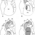
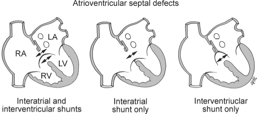
- – Atrioventricular septal defects (endocardial cushion defects) (Fig. 12.2)
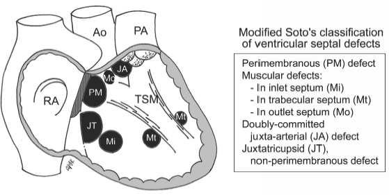
- – Ventricular septal defects (Fig. 12.3). Classified according to the location of the defect within the septum seen from the right ventricle and its relationships to the membranous septum, atrioventricular valves, and semilunar valves.
- ○ Perimembranous defects: the most common type and involves the membranous septum and adjacent muscular septum
- ○ Muscular defects: completely surrounded by the muscle
- ○ Doubly committed juxtaarterial defect: involves the most cranial part of the septum. The aortic and pulmonary valves are in fibrous continuity in the roof of the defect.
- ○ Non-perimembranous, juxtatricuspid defect: exceedingly rare defect involving the inlet part of the muscular ventricular septum along the tricuspid annulus
- • Extracardiac arterial
- – Ruptured sinus of Valsalva
- – Coronary artery-cameral fistulas
- – Anomalous coronary artery arising from the pulmonary artery
- – Aortopulmonary window
- – Patent ductus arteriosus
- • Extracardiac venous
- – Partial anomalous pulmonary venous connections
 Atrial, atrioventricular, and ventricular septal defects as well as patent ductus arteriosus, which constitute most left-to-right shunt lesions, are discussed below.
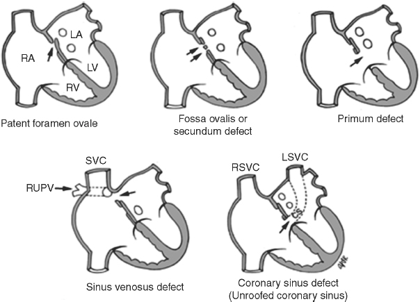
Fig. 12.1 Atrial septal defects. RUPV, right upper pulmonary vein; SVC, superior vena cava; CS, coronary sinus; LSVC, left superior vena cava; RSVC, right superior vena cava; RA, right atrium; LA, left atrium; RV, right ventricle; LV, left ventricle.
Fig. 12.2 Atrioventricular septal defects. LA, left atrium; LV, left ventricle; RA, right atrium; RV, right ventricle.
Fig. 12.3 Ventricular septal defects. RA, right atrium; Ao, ascending aorta; PA, pulmonary artery; TSM, trabecular septomarginalis.
Pathophysiology
 A spectrum of lesions with either an intracardiac or extracardiac communication whose fundamental abnormality is allowing blood to shunt from the higher pressure systemic circulation to the lower pressure pulmonic circulation, resulting in oxygenated blood recirculating through the lungs
 Levels of shunt
 Atrial, atrioventricular, and ventricular septal defects as well as patent ductus arteriosus, which constitute most left-to-right shunt lesions, are discussed below.


 Distinguished by different anatomic communications: The common denominator is that the pulmonary flow is greater than systemic flow.
 Coexisting cardiac lesions may significantly affect shunt volume.
 Hemodynamic severity varies according to the site of the defect.