Presentation and Presenting Images
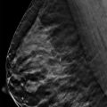
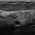
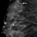
( ▶ Fig. 12.1, ▶ Fig. 12.2, ▶ Fig. 12.3, ▶ Fig. 12.4)
A 60-year-old female with history of right breast cancer treated with lumpectomy and radiation therapy presents for a screening mammogram.
12.2 Key images
12.2.1 Breast Tissue Density
There are scattered areas of fibroglandular density.
12.2.2 Imaging Findings
In the upper outer and the lower inner quadrants of the right breast, there are large linear rodlike calcifications. The calcifications in the lower inner quadrant are well seen in a ductal distribution on the DBT images ( ▶ Fig. 12.5 and ▶ Fig. 12.6).
12.3 BI-RADS Classification and Action
Category 2: Benign
12.4 Differential Diagnosis
Secretory calcifications: These calcifications are thick and rodlike in a ductal pattern radiating from the nipple.
Ductal carcinoma in situ (DCIS): DCIS and secretory calcifications can have the same distribution as they both involve the ducts. The calcifications in DCIS are often finer, irregular, and not smooth like those seen in secretory calcifications.
Vascular calcifications: In the early phase of vessel-wall calcification, the hallmark parallel ”tram-track” appearance may not be present. The calcifications are less dense and can appear as “dot-dash” and linear. These calcifications do not have this appearance.
12.5 Essential Facts
Secretory calcifications are often described as large rodlike. They are greater than or equal to 1 mm in diameter and can be 3 to 10 mm in length.
Secretory calcifications require no treatment or intervention, except for routine follow-up.
The large size of secretory calcifications makes them clearly delineated on DBT.
The orientation of the secretory calcifications in this case allow for large regions of the ductal distribution to be identified in both the craniocaudal (CC) and mediolateral oblique digital breast tomosynthesis (MLO DBT) image sets.
12.6 Management and Digital Breast Tomosynthesis Principles
Most secretory calcifications are detected on routine screening.
Secretory calcifications can increase over time.
DBT likely does not contribute much to making the diagnosis of secretory calcifications compared to full-field digital mammography (FFDM). It is important to be aware of the different patterns of benign calcifications on DBT.
When secretory calcifications are extensive, it can be difficult to identify suspicious calcifications in their midst.
The early phase of secretory calcifications can mimic DCIS.
12.7 Further Reading
[1] Baker JA, Lo JY. Breast tomosynthesis: state-of-the-art and review of the literature. Acad Radiol. 2011; 18(10): 1298‐1310 PubMed
[2] Spangler ML, Zuley ML, Sumkin JH, et al. Detection and classification of calcifications on digital breast tomosynthesis and 2D digital mammography: a comparison. AJR Am J Roentgenol. 2011; 196(2): 320‐324 PubMed

Fig. 12.1 Right craniocaudal (RCC) mammogram.
Stay updated, free articles. Join our Telegram channel

Full access? Get Clinical Tree


