Chapter 153
Lipoma
Epidemiology
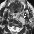
The parapharyngeal space is predominantly fat-filled, and primary tumors in this space are uncommon. The most common primary tumor is a pleomorphic adenoma. Occasionally, a lipoma may be seen in this space. Most of the lipomas found in the head and neck are located in the triangle.
Clinical Findings
Lipomas are more common in overweight individuals. They may show rapid increase in size during periods of weight gain. They often present as incidental findings on imaging but patients may also complain of vague neck discomfort. When these tumors are large enough, they may encroach on the adjacent spaces and present as mass lesions.
Pathology
Lipomas are yellow, lobulated, and very well encapsulated tumors. They consist of normal adult adipose tissue. Lipomas are classified histologically according to the kind of other tissues that may be present (e.g., fibrolipoma, angiolipoma, and myxolipoma). These tumors very rarely undergo malignant degeneration. Liposarcomas originate from lipoblasts within fascia rather than ordinary lipocytes.
Treatment