Localization: Anywhere there is fat. Most common in thigh, retroperitoneum, inguinal region, and popliteal fossa. Never in the hands, feet, and neck.
Clinical: Insidiously growing, deep-seated, ill-defined mass that usually reaches a large size. Pain, tenderness, and functional disturbances occur in 10–15 % of cases, but these are usually late complaints. Tumor may be painful due to compression on the nerve or cause edema of the limb due to venous occlusion. Retroperitoneal tumor may cause hydronephrosis, intestinal compression, inguinal hernia, and edema of the lower limbs.
Imaging: On X-ray: in more differentiate varieties, well-delineated radiolucency clearly distinguished from the surrounding muscles. Occasionally calcification, little vascularity. In more undifferentiated cases, less well-defined masses with greater density. On CT scan: in lipoma-like type, well-marginated mass mimicking lipoma. Frequently, thickened linear, nodular septa that enhance after contrast. In myxoid type, encapsulated, septated, slightly heterogeneous mass mimicking a cyst. Density of water. On MRI the myxoid type shows a homogeneous low signal intensity (dark) on T1 and high (white) on T2, strong enhancement, and minute foci of fatty tissue; small bright streaks on T1 in an overall dark lesion are typical. Nonspecific appearances are displayed by the other varieties: heterogeneous signal intensity on T1, white and dark areas on T2, and enhancement of all components. In dedifferentiated liposarcoma, a homogeneous mass of low signal intensity on T1 converts to a lesion with bright and intermediate areas on T2. Often the tumor is adherent or erodes adjacent cortical bone that responds with modest periosteal reaction. On bone scan liposarcomas are inordinately hot in contrast to other malignant soft tissue tumors. On angiography: richly vascular with intense intra- and peritumoral neoangiogenesis.
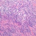
Histopathology: Usually very large, multilobulated, and delimited by a thin reactive tissue (pseudocapsule) interrupted by some tumor nodules. Satellites are present.
Varieties:
(a)
Well-Differentiated Liposarcoma/Atypical Lipomatous Tumor (WDL/ALT): 40 % of all cases.
1.
Liposarcoma lipoma-like: The most common. Yellowish, soft, oily, and friable. Mature fat mixed with collagen. Lipoblasts and lipocytes with hyperchromic nuclei and hyperchromic stromal cells within fibrous septa. Easy to confuse with lipoma.
2.
Sclerosing liposarcoma: More frequent in retroperitoneum. Whiter and firmer. Scattered bizarre stomal cells associated with multivacuolated lipoblasts. Abundant collagen. Easily confused with a scar.
3.
Inflammatory: More frequent in retroperitoneum. Like 1 and 2 admixed with extensive chronic inflammation. Easily mistaken for inflammatory process involving fat.
(b)
Myxoid (ML): Most frequent in the limbs (30–35 %). Soft, pale yellow, mucoid, and translucid or bright cherry red. Proliferating lipoblasts in different stages. A myxomatous background between cellular areas. Myxoid substance rich in mucopolysaccharides with a large quantity of hyaluronic acid. A network of thin capillaries of uniform caliber with an arborizing pattern. Oval to stellate cells scattered in the myxoid substance. Mitotic activity is rare. Hypercellular areas can be present showing ovoid-round, hyperchromic cells with scanty lipoblasts isolated or collected in small clusters. These are the features of a neoplasm that was formerly known as round cell liposarcoma. Myxoid and round cell liposarcoma represent a morphologic continuum of myxoid adipocytic neoplasia. This is supported by genetic analysis demonstrating the exact same characteristic chromosome translocations. The two main karyotypic aberrations are t(12;16) that fuses the DDIT3 gene on 12q13 with the FUS gene on 16p11 and t(12;22) that fuses DDIT3 with EWS on 22q12.
(c)


Pleomorphic (PL): Frequent (20 %). Softer, encephaloid, very cellular tissue with a nondescriptive pattern recognizable as sarcoma but with no unique features. On high power pleomorphism is prominent. Pleomorphic lipoblasts with vacuolated cytoplasm are scattered among undifferentiated hyperchromatic neoplastic cells. Large atypical, multinucleated giant cells are scattered through the tissue. Many atypical mitosis. S-100 immunoreactivity may help highlight the presence of multivacuolated lipoblasts in those cases in whom adipocytic differentiation tends to be focal and, therefore, easily overlooked. Similar to most pleomorphic high-grade sarcomas, pleomorphic liposarcomas tend to exhibit complex karyotypes.
Related posts:


Stay updated, free articles. Join our Telegram channel
Full access? Get Clinical Tree
