Location: Spinal roots, nerves of the mediastinum or of the retroperitoneum, peroneal, and ulnar nerve. Single nodule.
Clinical: Slow and progressive pain and neurological symptoms. Liquoral block, compression of the other roots or of the spinal cord, rachialgia, nocturnal increase in pain, stiffness, spinal contractures. Sharply painful, irradiated pain, paresthesia, algodystrophic syndrome when in peripheral nerve.
Imaging: On x-ray: scalloping of bone. On CT: well-circumscribed, homogeneous lesion, with muscle density. Frequent nonenhancing necrotic and cystic areas that cause an inhomogeneous hyperdense lesion after contrast administration. On MRI: nerve along the site of the mass. Capsulated. Homogeneous, isointense to muscle with frequent areas of low signal on T1, inhomogeneous, sometimes target appearance, higher than fat intensity on T2, diffused or peripheral enhancement with central necrotic unchanged zones on contrast T1.
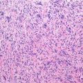
Histopathology: Globose “sausage-like” or “dumbbell mass,” elongated along the sac, well delimited by a capsule, soft, pink, white, or yellow, connected to a nerve root. Rarely, it expands like an hourglass inside and out of the intervertebral root foramen. S. of the peripheral nerves appears like an “onion bulb” or a piece of fruit attached to its stem. When very large, it may cause wide osteolyses. Usually encapsulated. Proliferation of spindle cells with ill-defined cytoplasm, in interlacing bundles, nuclear palisading, whorling of the cells, Verocay body formed by compact rows of well-aligned nuclei and cell processes that assume a roughly oval shape. Compact areas of spindle cells (Antoni A) alternating with loosely arranged areas (Antoni B). Rare if any mitotic figures. Large, irregularly spaced vessels with thickened wall and filled with thrombus material.
Course and Staging: Slow growth, rare recurrences after even incomplete surgery. Usually, stage 1 or 2.
Treatment: Marginal excision is curative. Malignant transformation exceptional.
Related posts:


Stay updated, free articles. Join our Telegram channel

Full access? Get Clinical Tree


