Localization: Skeletal muscles of extremities and retroperitoneum. Tumor is deep in 90 % of cases.
Clinical: Globose and painless swelling with no characteristic clinical features other than a frequently rapid growth rate.
Imaging: On X-ray: nonspecific STT displacing the adjacent fat. Peripheral calcifications are rare (9 %). Florid periosteal reaction and smooth cortical erosion may be observed. In these cases, bone scan is always very hot. On angiography: typical changes occurring in sarcomas, with very large avascular areas of tumor due to necrosis or hemorrhage. Major vessels are almost never infiltrated. On CT: inhomogeneous, similar to or lower density than that of muscle, strong enhancement of the solid component, central hypodense area of necrosis, hemorrhage, myxomatous tissue, with large cavities with fluid contents, and a thick wall that are mistaken with a hematoma. On MRI: poorly defined margins, homogeneous, muscular intensity on T1 and heterogeneous high signal intensity on T2, dark central necrotic zones and strong enhancement at the periphery on contrast T1, internal low signal intensity septa of collagen bands on T1 and T2. In MFH, central myxoid area is black on T1 and white on T2. Hematoma is white on T1; fluid levels show low signal intensity for hemosiderin deposits and high for supernate on both sequences.
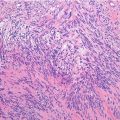
Histopathology: White-gray mass with no distinctive macroscopic features other than the frequent presence of necrosis. Pleomorphic aspect, cellular atypia with hyperchromic nuclei, coarse chromatin, large nucleoli, and numerous typical and atypical mitotic figures. Histologically, UPS resembles other specific type of pleomorphic sarcoma, with frequent multinucleated tumor giant cells and a frequent patternless pattern. In some areas, there is a distinctive orientation around eosinophilic areas or vessels that gives the appearance of a cartwheel: “the storiform pattern.” Collagen production may produce an accentuation of this feature. Clusters of histiocytes, foam cells, and inflammatory cells are sprinkled. In the 2013 WHO classification, UPS is classified in the group of undifferentiated/unclassified sarcomas that are divided into pleomorphic (UPS), round cell (similar to other specific types of round cell sarcoma, especially Ewing’s sarcoma), spindle cells, and epithelioid (similar to a metastatic carcinoma or melanoma) subsets. On immunohistochemistry, undifferentiated pleomorphic sarcomas are positive for vimentin, and by definition, no pattern of protein expression that would identify a specific line of differentiation can be identified.
Related posts:


Stay updated, free articles. Join our Telegram channel

Full access? Get Clinical Tree


