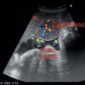
Liver is divided into left and right lobe and various segments depending on the vascular supply. It has a dual blood supply from the portal vein and the hepatic artery, both directing the blood toward the liver. Three hepatic veins drain the blood from the liver into IVC, exhibiting a trident appearance on USG.
Left hepatic vein: Divides left lobe into medial and lateral segments.
Middle hepatic vein: Separates left and right hepatic lobe, also separated by gallbladder (GB) fossa; courses within the main lobar fissure.
Right hepatic vein: Divides right lobe into anterior and posterior segments.
Quadrate lobe: Medial segment of left hepatic lobe. Located between round ligament and GB fossa.
Caudate lobe: First segment of liver; bounded anteriorly by ligamentum venosum and posteriorly by IVC. It has its own blood supply and drainage. Should not be mistaken for a lymph node.
Glisson’s capsule: Surrounds the liver and is thickest around the porta hepatis and IVC.
Main Portal vein: Divides into right and left portal vein.
Right portal vein has anterior and posterior division.
Left portal vein has medial and lateral divisions.
They supply blood to the liver in their related segments (Figure 2.1 and Table 2.1).
Porta hepatis: Site at which portal vein, common bile duct (CBD), and hepatic artery are located within peritoneal fold called the hepatoduodenal ligament.
In utero, umbilical vein in liver divides into left and right.
Left umbilical vein: Connects directly to left portal vein. After birth, it becomes fibrous cord (Ligamentum teres/Round ligament). Ascends along the falciform ligament. Divides left lobe into medial and lateral segments.
Right umbilical vein (Ductus Venosus): Shunts blood directly into the IVC. After birth, it collapses and becomes ligamentum venosum.
Figure 2.1 Illustrates segmental anatomy of liver.
Table 2.1 Illustrates Couinaud’s segmental anatomy of liver
Segment 1 | Caudate lobe |
Segment 2 | Lateral segment left lobe (superior) |
Segment 3 | Lateral segment left lobe (inferior) |
Segment 4 (A&B) | Medial segment left lobe |
Segment 5 | Anterior segment right lobe (inferior) |
Segment 6 | Posterior segment right lobe (inferior) |
Segment 7 | Posterior segment right lobe (superior) |
Segment 8 | Anterior segment right lobe (superior) |
Coronary ligament: Right layer of the falciform ligament
Left triangular ligament: Left layer of the falciform ligament
Right triangular ligament: Most lateral portion of the coronary ligament
Bare area: The posterosuperior region of the liver, which is not covered by peritoneum
Usually convex transducer (3.5 MHz) is used.
Nil by mouth except water for 8 hours before examination in adults and two to 3 hours in infants. In acute cases, immediate scanning may be required.
Patient should be scanned in supine, left oblique, and left lateral decubitus positions.
Patient should be scanned in axial, sagittal, and oblique planes, including subcostal and intercostal scanning.
1. Hepatomegaly
2. Right upper quadrant (RUQ) pain
3. Jaundice
4. Suspected liver abscess
5. Suspected liver mass and metastasis
6. Ascites
7. Trauma
Size: Usually <15 centimeters in midclavicular line.
Eye-balling technique: Liver extending below the lower pole of the right kidney is suggestive of hepatomegaly.
Reidel’s lobe: Extension of the right lobe of liver, which is a normal variant.
Beaver tail liver: Sliver of liver, a variant where an elongated left lobe surrounds the spleen.
Echogenicity of Pancreas > Liver ~ Spleen > Kidneys (cortex > medulla)
Homogenous parenchyma with thin-walled hepatic veins and bright reflective portal veins (Figure 2.2)
Figure 2.2 Depicting bright reflective portal vein walls and thin nonreflective hepatic vein walls.
Hepatitis → Inflammation of liver due to varying etiologies.
• Liver may appear normal.
• Tender hepatomegaly.
• Diffusely reduced/altered echogenicity.
• Increased brightness of portal triad (Periportal cuffing)—Starry sky pattern (Figure 2.3).
• Thickened edematous GB wall.
Liver may appear normal or reveal coarse echotexture with reduced echogenicity and diminished brightness of portal triad.
End-stage parenchymal disease.
• Coarse echo pattern secondary to nodularity and fibrosis. Micro- (<1 centimeter) and macronodules (up to 5 centimeters) may be seen
• Hepatomegaly in early stages and shrunken liver in advanced cases (Figure 2.4)
• Ascites
• Enlarged caudate lobe (width of C/RL-caudate lobe/right lobe >0.65 is suggestive of (S/o) cirrhosis)
• Splenomegaly
• Dilated portal vein
• Reduced vascular markings with decreased attenuation
Figure 2.3
Stay updated, free articles. Join our Telegram channel

Full access? Get Clinical Tree


