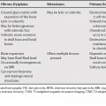
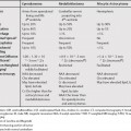
70 The most common lesions with a central scar are focal nodular hyperplasia (FNH), fibrolamellar hepatocellular carcinoma (HCC), and hemangiomas. Hemangiomas can usually be differentiated by the characteristic enhancement pattern. FNH and fibrolamellar HCC can be more difficult to differentiate as they are both hypervascular tumors with central scars seen in young patients. Many imaging features can be helpful in differentiation of these two lesions (Table 70.1).
Liver Lesions with Central Scar
| FNH | Fibrolamellar HCC | |
|---|---|---|
| Sex | Female | No sex predilection |
| Size >5 cm | – | + |
| Surface Lobulation | – | + |
| Heterogeneity | – | + |
| Calcifications | – | ± |
| Capsular Retraction | – | Rare |
| Scar Size | <2 cm | >2 cm |
| Scar Intensity | T2WI hyperintense | T2WI hypointense |
| Delayed Scar Enhancement | + | – |
| Capsule* | – | ± |
| Pseudocapsule† | ± | ± |


