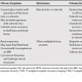
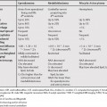
69 Lesions with delayed enhancement have prominent enhancement occurring after the arterial phase. Delayed enhancement in a liver lesion usually corresponds to fibrotic tissue.1 Some lesions (e.g., cavernous hemangiomas) have prolonged enhancement. This is early enhancement that persists on delayed images. Capsular enhancement in hepatocellular carcinoma (HCC) can be delayed or prolonged.2 Central prolonged enhancement in HCC is rare, but is sometimes seen in scirrhous HCCs. (Due to the higher contrast resolution of magnetic resonance imaging (MRI), this is seen more often on MRI than on computed tomography [CT].)3 Delayed enhancement in metastases is most commonly seen in metastatic colorectal adenocarcinoma. This delayed enhancement occurs in the central portion of the lesion.2 Confluent hepatic fibrosis is seen in advanced cirrhosis. It is wedge-shaped and usually involves the anterior and medial segments. It is associated with capsular retraction.4
Liver Lesions with Delayed/Prolonged Enhancement
Features of Specific Lesions with Delayed/Prolonged Enhancement
Hepatocellular Carcinoma
Metastases
Confluent Hepatic Fibrosis
Inflammatory Pseudotumor
Related posts:

Stay updated, free articles. Join our Telegram channel

Full access? Get Clinical Tree


