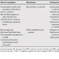
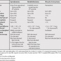
76 Hyperintensity on T1-weighted magnetic resonance images (T1WI) in a liver lesion could be secondary to macroscopic fat or hemorrhage, but some hepatic lesions can be hyperintense without hemorrhage or macroscopic fat. This could be secondary to microscopic fat, copper, protein, mucin, or melanin.1,2 Regenerative nodules in cirrhosis are usually hypointense on T1WI (unlike dysplastic nodules, which are typically hyperintense). Rarely, regenerative nodules can be hyperintense.3 Benign regenerative hepatic nodules have been described in Budd-Chiari syndrome as multiple, small, and hypervascular. These are likely a result of insufficient blood supply to portions of the liver, which will atrophy, with compensatory nodular hyperplasia in areas of the liver with adequate blood supply. These nodules have also been reported in other systemic disorders that impair hepatic blood flow, such as autoimmune disease and lymphoproliferative and myeloproliferative disorders; they can occur after treatment with steroids and antineoplastic medications as well.4 Similar nodules have also been described in autoimmune hepatitis.5 These regenerative nodules are distinct from nodular regenerative hyperplasia (see Chapter 73),6
Liver Lesions with Hyperintensity on T1-Weighted Magnetic Resonance Imaging
![]()
Stay updated, free articles. Join our Telegram channel

Full access? Get Clinical Tree


