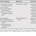
77 Liver lesions can contain areas of T2-weighted magnetic resonance imaging (T2WI) hypointensity secondary to hemorrhage or calcification. Other etiologies include fibrosis, desmoplastic stroma, cellular necrosis, mucin, and iron deposition.1,2 Lesions can be diffusely hypointense (e.g., a siderotic regenerating nodule), have heterogeneous areas of hypointensity (e.g., from hemorrhage), or central hypointensity (e.g., a fibrotic central scar in cholangiocarcinoma).
Liver Lesions with Hypointensity on T2-Weighted Magnetic Resonance Imaging
Liver Lesions with Hypointensity on T2WI2–7
Related posts:

Stay updated, free articles. Join our Telegram channel

Full access? Get Clinical Tree


