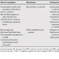
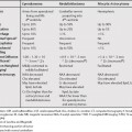
74 The most common solitary tumors containing fat are hepatocellular carcinoma and hepatic adenoma.1 Fatty change in adenomatosis is more frequent compared with a solitary adenoma. Macroscopic fat in a focal nodular hyperplasia is very rare.2 Metastases that contain macroscopic fat are rare, but include metastatic ovarian dermoid, teratoma, liposarcoma, and Wilms’ tumor.3 A less common lesion with macroscopic fat is hepatic angiomyolipoma.3,4 Angiomyolipomas are rare benign hepatic tumors, which are found predominately in women. They have been associated with tuberous sclerosis and renal angiomyolipomas. Three patterns can be seen on imaging: approximately equal portions of fat, vessels, and smooth muscle; a predominance of fat component, and a minimal fat component. Angiomyolipomas with approximately equal portions of fat, vessels, and smooth muscle have the most characteristic appearance. Angiomyolipomas with a predominance of fat are usually not distinguishable from myelolipomas or lipomas. Lesions with minimal fat are often difficult to distinguish from hepatocellular carcinoma (HCC) with fatty change. Both angiomyolipomas with minimal fat and HCCs with fatty change can appear as hypervascular masses with areas of fat. Numerous features are helpful in distinguishing the two lesions (Table 74.1).
Liver Lesions with Macroscopic Fat
Stay updated, free articles. Join our Telegram channel

Full access? Get Clinical Tree


