Liver: Portal Venous System and Hepatic Veins


10.1055/b-0034-87862

Liver: Portal Venous System and Hepatic Veins


Table 2.6 Abnormal porta hepatis

Diagnosis


Findings


Comments


Pseudotumor caused by bowel loops


US: multiple attempts at visualization and peristalsis usually suffice.


Repeat examination after fluid administration is useful. CT with oral contrast is rarely necessary.


Periportal lymphadenopathy


Multiple solid nodes of different sizes, sometimes with central vascularization.


Burkitt lymphoma is the most frequent cause.


Cavernous transformation of the portal vein


Fig. 2.18, p. 154


Best demonstrated by color Doppler US and MR angiography.


US shows a mass of tubular structures, within or around a previously thrombosed portal vein.


Doppler US: the porta is replaced by a tangle of venous collaterals at the hepatic hilum whose purpose is to link the extrahepatic portal circulation and the intrahepatic portal branches.


Extrahepatic and intrahepatic portal vein thrombosis


US: Direct visualization of the thrombus is rare in children. The portal vein usually appears normal or the portal lumen is absent. Doppler is essential as it demonstrates absence of blood flow. Occasionally, there can be total absence of flow in the hepatic hilum, with reversed portal flow in peripheral intrahepatic branches (spontaneous arterioportal shunts).


Chronic thrombosis leads to cavernomatous transformation of the portal vein.


Causes: portal vein thrombosis (perinatal omphalitis or umbilical venous catheterization, sepsis, appendicitis, acute dehydration, coagulopathies, splenectomy, hepatic transplantation, cirrhosis related to biliary atresia, congenital hepatic fibrosis).


Portal vein thrombosis leads to portal hypertension, manifested by the presence of collateral circulation, possible recanalization of the periumbilical veins, less omental thickening, or splenomegaly.

Fig. 2.18 Cavernous transformation of the portal vein. Oblique US through the hepatic hilum shows multiple tubular structures without evidence of normal portal vein.


Table 2.7 Abnormal periportal spaces

Diagnosis


Findings


Comments


Posttraumatic


A harbinger of other injury in the peritoneal cavity.


Post liver transplant


Due to lymphatic stasis.


Leukemia and lymphoma


US: Markedly enlarged and echogenic liver. Widened periportal areas, predominantly hypoechoic.


Periportal tumoral infiltration.


Cirrhosis


Bandlike echogenic, periportal areas. A rare finding in cirrhosis.


Fatty liver


Echogenic changes in the periportal regions seen in marked obesity.


Periportal fibrosis


Sequela of previous disease, including cytomegalovirus hepatitis, alpha1-antitrypsin deficiency, post liver transplant, vascular abnormalities.


Niemann-Pick disease


Histological diagnosis: deposition of sphingomyelin.


Bile Ducts and Gallbladder


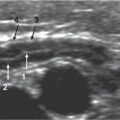
Ultrasound (US) is the imaging modality of choice for screening the gallbladder and biliary tract in children. The study should consist of a systematic examination of the liver, bile ducts, gallbladder, pancreas, and spleen. A complete duplex and color Doppler examination of the hepatic vessels should be performed. US guidance is very useful to guide invasive diagnostic or therapeutic procedures.


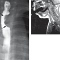
Magnetic resonance cholangiopancreatography (MRCP) is also a noninvasive, nonradiating technique; in young children, MRCP needs to be performed under sedation. MRCP uses the signal from the bile within the ducts to allow visualization of the biliary system. Fasting is necessary. The basic principle is to use a heavily T2-weighted sequence with fat suppression to show the high signal intensity of the static fluids. MRCP has the potential to replace diagnostic endoscopic retrograde pancreatography (ERCP) and percutaneous transhepatic cholangiography. A normal study obviates the need for more invasive studies.


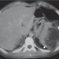
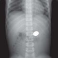
Computed tomography (CT) has very few indications because of the use of radiation and, frequently, intravenous contrast. CT is useful in differentiating aerobilia from calculus. Multidetector equipment allows fast scans and three-dimensional (3D) reconstruction of very good quality even in non–breath-holding children.


ERCP is invasive and technically diffcult. It requires general anesthesia in children and has the potential for significant associated morbidity. However, it is a useful diagnostic and therapeutic tool in selected cases.


Hepatobiliary scintigraphy is an isotopic study that uses technetium (99mTc)-labeled iminodiacetic acid that is extracted and secreted by the liver. Scintigraphic visualization of tracer in the gastrointestinal (GI) tract indicates patency of the extra hepatic biliary ducts. It is useful to demonstrate bile leakage after surgery and for diagnosing of biliary atresia.


Bile Ducts


Table 2.8 Dilated bile ducts

Diagnosis


Findings


Comments


Caroli disease


Fig. 2.19


US findings: dilated, ectatic bile ducts, “intraluminal portal vein” or “central dot” sign with flow on Doppler, biliary sludge and calculi, enlarged gallbladder and common bile duct, abscesses, and associated renal anomalies.


Recognition of the connection of the ectatic ducts with one another and with the rest of the ductal system is critical (DD between Caroli disease and polycystic disease).


Segmental nonobstructive dilatation of the intrahepatic bile ducts. Ductal plate malformation spectrum. It is associated with hepatic fibrosis, choledochal cyst, medullary sponge kidney, infantile polycystic renal disease, or nephronophthisis.


Ascending cholangitis


Associated with biliary obstruction whether congenital or acquired.


Appropriate therapy has to be directed to the underlying cause.


Sclerosing cholangitis


Strictures in multiple segments of the bile ducts with intervening dilated segments. Amputation of some segmental bile ducts is common.


MRCP is the first imaging modality (demonstrates the typical abnormalities with high specificity).


Inflammatory obliterative fibrosis affecting the intraand extrahepatic biliary tree. Poor prognosis with progressive evolution to biliary cirrhosis.


Choledochal cyst


Fig. 2.20, p. 156


US: Cystic dilatation of the common bile duct often associated with hepatic bile duct dilatation. (This affects the main left and right hepatic ducts but not the smaller ducts.)


MRCP provides the anatomic map of the pancreaticobiliary ductal union and also of the biliary tree, which are critical for surgery.


Congenital. Most frequent cause of extrahepatic cholestasis in childhood.


Bile plug syndrome


Sludge in the bile ducts and gallbladder forms plugs and may cause transient gallbladder and duct dilatation in the neonatal period.


Choledocholithiasis


Fig. 2.21a, b, p. 156


Fig. 2.22a, b, p. 156


US: echogenic material within the dilated duct with associated acoustic shadowing.


Most frequent cause of biliary obstruction.


Biliary obstruction without calculi


DD with cavenous transformation of the portal vein by Doppler US.


Caused by biliary tumors (rhabdomyosarcoma of the biliary tree), inspissated bile, pancreatitis, congenital biliary stenosis.


Biliary obstruction in liver transplantation


US: dilated intrahepatic ducts with dilatation of the proximal common bile duct to the level of the surgical anastomosis.


MR cholangiography can be used to delineate the anatomy and morphology of bile ducts and to search for biliary strictures.


Children with reduced-size transplantation have a higher risk. Most anastomotic strictures are secondary to scar tissue causing retraction and narrowing of the common bile duct at the suture site, although ischemia may also be a factor.


Segmental saccular dilatations in transplanted livers are mostly secondary to arterial thrombosis or stenosis.

Fig. 2.19 Caroli disease. Doppler US of the liver shows an avascular cysticlike structure with surrounding portal vessels that correspond to an intrahepatic biliary duct.
Fig. 2.20 Choledochal cyst. Transverse US image depicts a cystic dilatation of the common biliary duct at the hepatic hilum (between calipers) with lithiasis inside the lumen.
Fig. 2.21a, b Choledocholithiasis. (a) Transverse and (b) longitudinal US scans showing a hyperechoic biliary stone with posterior shadowing located in the common bile duct. The cystic duct is also dilated in the longitudinal image.
Fig. 2.22a, b Choledocholithiasis. (a) MRCP shows an obstructing material in the distal common bile duct and the biliary and pancreatic ducts dilatation. (b) Percutaneous cholecystography also demonstrates the stone producing the bile duct dilatation.


Table 2.9 Abnormal nondilated bile ducts

Diagnosis


Findings


Comments


Aerobilia


DD: portal venous air in necrotizing enterocolitis.


Abdominal radiographs and Doppler US are the best imaging modalities for diagnosing aerobilia.


After biliary tract surgery or manipulation, as well with incompetent ampulla of Vater; in biliary atresia, after Kasai procedure; in reduced-size liver transplant (choledochojejunostomy).


Sclerosing cholangitis


Irregular segmentary narrowing of the bile ducts with cholestasis. Secondary pancreatitis may occur. Diagnosis is made on liver biopsy.


Associated with ulcerative colitis, Crohn disease, autoimmune hepatitis, or idiopathic causes.


Bile duct tumors


Papilloma and adenoma.

Only gold members can continue reading. Log In or Register to continue

Stay updated, free articles. Join our Telegram channel

Jul 12, 2020 | Posted by in PEDIATRIC IMAGING | Comments Off on Liver: Portal Venous System and Hepatic Veins

Full access? Get Clinical Tree

Get Clinical Tree app for offline access