The Gastrointestinal Tract: Esophagus


10.1055/b-0034-87865

The Gastrointestinal Tract: Esophagus


Displacement and Compression


Esophageal displacement can be demonstrated by means of an upper GI (UGI) study, MRI, or CT.


Table 2.30 Esophageal displacement

Diagnosis


Findings


Comments


Neurogenic tumors (neuroblastoma, neurofibroma)


Solid homogeneous masses in the posterior mediastinum. Anterolateral displacement of the esophagus. May calcify.


May produce osseous involvement. Additional investigation: MRI, (123) I-metaiodobenzylguanidine [(123) I-MIBG] scintigraphy, biopsy.


Anterior intrathoracic meningocele


Fluid-filled soft-tissue density tumor in the posterior mediastinum. Dysraphic changes are common.


Often associated with neurofibromatosis. Additional imaging: CT, MRI.


Esophageal duplication


Fig. 2.58a, b, p. 180


Fig. 2.59a, b, p. 180


(see Table 2.34 )


May be associated with dysraphic vertebral bodies (neurenteric cyst) and other bowel duplications.


Abscess


Displacement by a spindle-shaped fluid collection on both sides of the vertebral bodies.


Descended retropharyngeal abscesses or secondary to spondylitis and discitis


Paraesophageal hernia


Lateral/anterior displacement of the distal esophagus.


Commonly filled with air. Either congenital or posthiatal hernia repair.


Cardiomegaly


Posterior displacement of the esophagus.


See Chapter 1


Aberrant vessels


See Chapter 1


Enlarged lymph nodes


Multiple solid homogeneous masses often in anterior and/or middle mediastinum.


Most often due to lymphoma.


Bronchogenic cysts


Cystic homogeneous lesion frequently in the middle mediastinum compressing the esophagus according to the size.


Arise from disruption in fetal separation of the esophagus and trachea. Work-up: CT, MRI.


Teratoma


Solid or cystic usually anterior mediastinal masses.


May contain bony elements and calcifications.


Work-up: CT, MRI.


Enlarged thymus


Posterior proximal esophageal displacement.


Rarely associated with swallowing or breathing diffculties.


Cystic lymphangioma


Cystic multiseptated mass with anterior mediastinum widening and causing posterior esophageal displacement.


The mass may extend from the cervical area through the thoracic inlet to the anterior mediastinum.


Pulmonary volume loss


Lateral displacement of the esophagus and the entire mediastinum to the side of the volume loss. Compensatory emphysema of the normal side.


Pulmonary aplasia, pulmonary volume loss/atelectasis, total lung atelectasis (foreign body), status postpulmonary resection.


Increased hemithorax volume


Lateral mediastinal displacement toward the uninvolved side.


Congenital lobar emphysema, congenital pulmonary cysts, cystic adenomatoid malformation, tension pneumothorax, diaphragmatic hernia.


Table 2.31 The esophagus: Dilatation and stricture

Diagnosis


Findings


Comments


Physiologic


Distention of esophagus during deglutition.


Due to swallowing of air or belching. In contrast to adults, gas in the esophagus is a frequent finding in children.


Hypotonia


Fig. 2.48


Generalized esophagus distention.


Secondary to inflammation and gastroesophageal reflux (GER).


GER (incompetent lower esophageal sphincter)


Fig. 2.49


Fig. 2.50


US or contrast examination show passage of gastric content to the esophagus.


Physiologic up to 18 mo of age. Endoscopic Ph-measure is the gold standard for diagnosis. Imaging is essential to rule out GER secondary to gastric outlet obstruction. Is the most frequent cause of aspiration pneumonia. Roviralta syndrome: hiatal hernia secondary to hypertrophic pyloric stenosis (HPS).
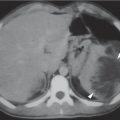
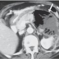

Hiatal hernia


Fig. 2.51


Anteroposterior X-ray: basal lucency within the outline of the cardiac silhouette.


Confirmation with contrast examination: to demonstrate GER and its sequelae (esophagitis and reflux stricture).


Mostly congenital. Sliding hernias varying in size. DD: small epiphrenic ampulla.


Reflux esophagitis


Fig. 2.52


Contrast examination: irregular and gross mucosal folds in distal esophagus with ulcerations.


The most important cause is hiatal hernia. Endoscopy for diagnosis.


Caustic injury


The mucosa is initially edematous and thickened.


Later, atonic dilatation occurs, as well as irregular mucosal pattern due to necrosis.


Nonionic contrast medium for diagnosis at early stage due to perforation risk. Late sequelae: long stenosis.


Most often with alkalis.


Other esophagitis (mycotic, viral, bacterial)


Spasm, pseudodiverticula ulcerations, edema, cobblestone pattern, stenosis.


Can affect the esophagus at any level.


Usually diffuse. Immunocompromised patients.
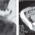

Foreign bodies


Fig. 2.53


Contrast examination: filling defects.


Foreign body impaction often at stenotic areas.


Barium or nonionic contrast medium in cases of perforation suspicion.


Endoscopic extraction.


Achalasia


Atonic esophagus dilatation (megaesophagus).


Heterogeneous air-fluid level due to air and retained food. The stomach bubble is small or absent. Contrast examination: characteristic beak deformity at the distal esophagus.


Rare in infants. Unknown cause.


Clinically: swallowing diffculty and retrosternal pain. DD: distal esophageal strictures.


Congenital esophageal stricture


Membranous-, hour-glass–, or tubular-shaped.


Associated with tracheoesophageal fistula or isolated as a cartilaginous ring in bronchial remnant syndromes.


Secondary esophageal stricture


Fig. 2.52, p. 175


Narrowed lumen with possible prestenotic dilatation. The contracture and scarring are more often in the middle and lower esophagus.


Causes: Most often due to reflux esophagitis. After operative repair for esophageal atresia, at the level of the anastomosis, after caustic esophagitis (alkalis, acids), with epidermolysis bullosa; postirradiation; with vascular anomalies.


Radiation damage


Mural scar formation and distortion with loss of motility. The lumen is narrowed.


Extent of the damage depends on the size of the radiation field.


Leukemic infiltration


Narrowing of the lumen, mostly in the distal esophagus.


Other tumors are rare.


Epidermolysis bullosa dystrophica


Circumferential constriction can occur, as well as long segment strictures.


Hereditary. Presents in infancy. Minimal trauma can result in blister production.


Scleroderma, dermatomyositis, lupus erythematosus


Dilatation. Absent peristalsis.


Hiatus hernia and reflux esophagitis may develop distal stenosis.

Fig. 2.48 Hypotonia. Plain anteroposterior chest radiograph shows a dilated hypotonic esophagus in a patient with GER.
Fig. 2.49 Gastroesophageal reflux. US sagittal view of an infant with GER: the lower esophageal sphincter is open and a large amount of gastric content is seen passing into the distal esophagus (arrowheads).
Fig. 2.50 Gastroesophageal reflux. Upper gastrointestinal (UGI) series in supine position: barium is seen refluxing into the esophageal hiatus to the lower esophagus.
Fig. 2.51 Hiatal hernia. UGI series in an infant: the barium-filled stomach is partially herniated and has an hourglass configuration.
Fig. 2.52 Peptic acid esophagitis. UGI series of an infant with esophageal stricture with some tiny lineal ulcerations caused by prolonged GER and peptic acid esophagitis.
Fig. 2.53 Foreign body. Plain chest film shows a radiopaque foreign body (coin) impacted in the upper third of the esophagus.

Only gold members can continue reading. Log In or Register to continue

Stay updated, free articles. Join our Telegram channel

Jul 12, 2020 | Posted by in PEDIATRIC IMAGING | Comments Off on The Gastrointestinal Tract: Esophagus

Full access? Get Clinical Tree

Get Clinical Tree app for offline access