Key Points
- ▪
Radiographically apparent aortic, mitral, tricuspid, and pulmonic valve prostheses can all be identified and localized by chest radiography. Older mechanical and biological valves were easy to identify because they generally had obvious metallic components. Contemporary mechanical and biological valves have far less or no ferrometallic or other metallic components; therefore, they are more difficult to recognize, especially if the heart shadow is dense. Furthermore, the momentum of prosthetic valve insertion has shifted toward bioprostheses (4:1 ratio), several of which have no radiographically evident components at all.
- ▪
The frontal radiograph alone is often adequate to identify and to localize a prosthesis, but superimposition of the shadow of a prosthesis onto that of the spine and obscuring of a prosthesis shadow by that of a large heart reduce the sensitivity of the frontal radiograph somewhat.
- ▪
The lateral radiograph is invaluable to allow projection of the prosthesis shadow off that of the spine.
- ▪
Both the position of the prosthesis shadow and its orientation are used to establish its location.
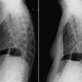
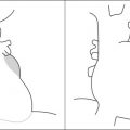
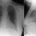
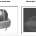
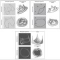
The chest radiograph can localize the great majority of valve prostheses to the aortic, mitral, or tricuspid positions, and it can usually determine whether the prosthesis is mechanical or bioprosthetic. In addition, the chest radiograph can offer radiographic evidence of the presence or absence of congestive heart failure. Both the posteroanterior and lateral radiographs are required ( Graphic 8-1 ).


Stay updated, free articles. Join our Telegram channel

Full access? Get Clinical Tree


