Michael Bret Anderson
Lower gastrointestinal (GI) bleeding is defined by intraluminal bleeding distal to the ligament of Treitz (the suspensory ligament of the duodenum composed of a band of smooth muscle extending from the junction of the duodenum and jejunum to the left crus of the diaphragm). The severity of lower GI bleeding can range from clinically insignificant to massive and life threatening. Although upper GI bleeding is four times as common as lower GI bleeding, lower tract bleeding still accounts for 21 adult hospital admissions per 100,000 persons per year. Primarily affecting older adults, the mean age of presentation is 63 to 77 years. Approximately 80% to 90% of cases will resolve with conservative medical therapy; however, up to 25% of patients will rebleed during or after the hospital admission. The mortality of lower GI bleeding is reported from 2% to 4%.1
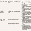
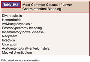
Common etiologies of lower GI hemorrhage in the adult population include diverticular disease, hemorrhoids, arteriovenous malformation (AVM) and angiodysplasia, postpolypectomy bleeding, inflammatory bowel disease, neoplasm, infection, ulcers, and aortoenteric fistula.2 Meckel diverticulum is encountered in the pediatric and young adult population (Table 30.1).
Lower tract varices are an uncommon source of lower GI bleeding and are generally treated with a similar approach to the more typical upper tract varices of portal hypertension.3
The colon is the source of bleeding in 80% of lower GI tract hemorrhages. When the source of bleeding is the large bowel, the distribution is rather evenly distributed, with one-third originating in the ascending colon, one-third in the transverse colon, and one-third in the rectosigmoid.4
Endoscopy is generally considered first-line treatment for both upper and lower GI bleeding.5 Although colonoscopy is useful and recommended for the evaluation and possibly the treatment of “slow” or chronic recurrent bleeding, its usefulness can be limited in the setting of acute lower GI arterial bleeding. Intraluminal blood and retained stool can impair visibility and make it difficult to reach the ascending colon, which is a common site of lower GI hemorrhage. Additionally, the lower GI tract to include all the small bowel distal to the ligament of Treitz cannot be fully evaluated with conventional endoscopy.1
Surgery, alternatively, does not carry the same limitations as endoscopy. However, emergent surgery for active arterial bleeding in the lower tract is associated with significant morbidity and mortality (20% to 30%), with mortality reported greater than 40% in some studies.6,7 In the event the site of bleeding cannot be localized intraoperatively, empiric resection might be required with selection of a partial or subtotal colectomy. If in fact the site of bleeding is not resected, hemorrhage is most likely to recur.8
The use of catheter angiography to diagnose GI bleeding was first described in 1963.9 The infusion of vasoconstrictive drugs followed soon thereafter. Then in 1970, autologous clot was first used as an embolic agent in the treatment of upper GI bleeding.10
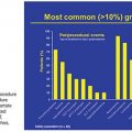
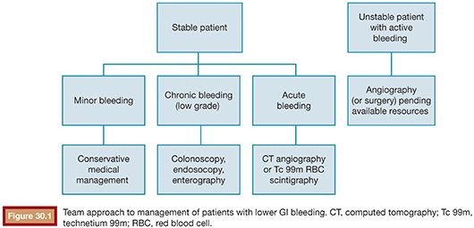
For maximum benefit and optimal outcomes, a multidisciplinary approach should be considered. A team including gastroenterology, interventional radiology, and surgery is recommended to provide the best care to the patient with lower GI bleeding (Fig. 30.1).
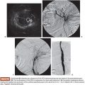
IMAGING (FIGS. 30.2 TO 30.8)
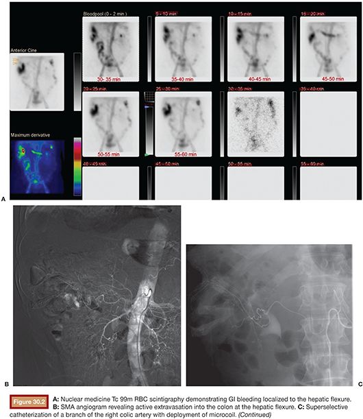
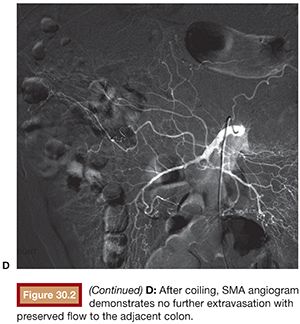
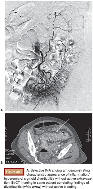
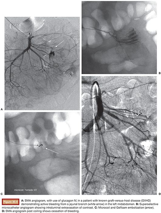
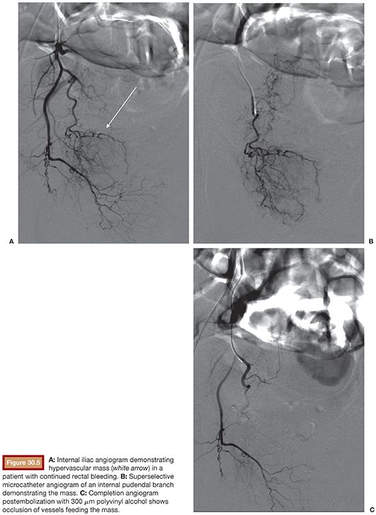
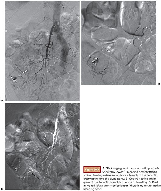
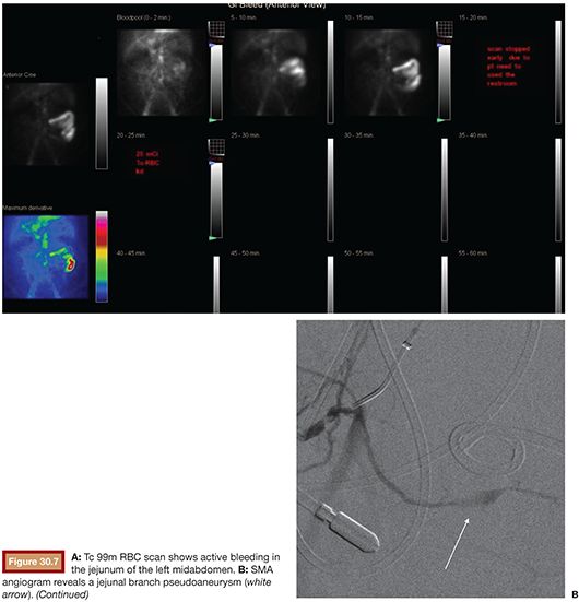
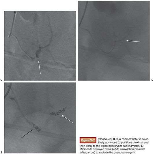
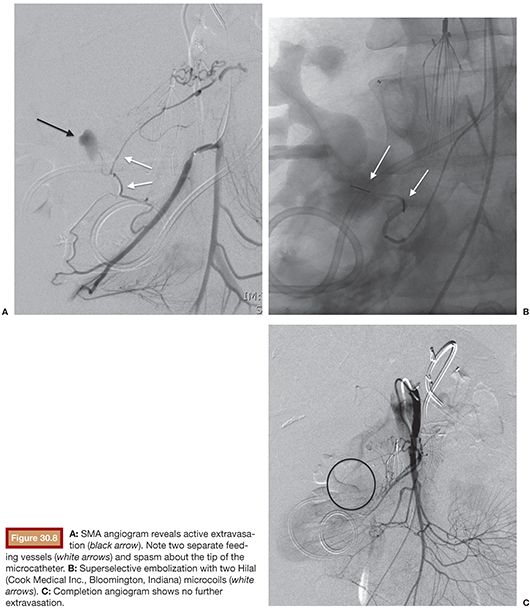
Given its sensitivity to detect GI bleeding at rates as low as 0.04 mL per minute, nuclear medicine radionuclide scintigraphy (“bleeding scan”) with technetium 99m (Tc 99m)–labeled red blood cells (RBCs) is often used to detect and localize GI hemorrhage. Tc 99m RBC scintigraphy has a reported 93% sensitivity and 95% specificity for detecting the site of GI bleeding.11 Scintigraphy is also of benefit in the patient demonstrating a pattern of recurrent hemorrhage who intermittently ceases to bleed. Because imaging is performed over the course of at least 1 to 2 hours, Tc 99m RBC studies increase the chances of “catching” active bleeding and therefore localizing the site before intervention.
However, in recent years, with the evolution of multidetector-row helical computed tomography (MDCT), contrast-enhanced computed tomography angiography (CTA) has become a rapid, noninvasive, and accurate tool to diagnose and localize GI bleeding. CT mesenteric angiography has been shown, in the setting of hemodynamically significant GI bleeding, to provide an overall location-based sensitivity, specificity, and accuracy in the detection and localization of GI bleeding of 91%, 99%, and 97%, respectively.12 In addition, CT provides the anatomic detail and diagnostic information of cross-sectional imaging that is not obtained with scintigraphy or catheter angiography alone. When the underlying etiology of bleeding is unknown, CT is very helpful in diagnosis. Given the multiple possible etiologies of lower GI hemorrhage, CTA can potentially limit the time and contrast volume that will be required in the angiography suite to localize and treat the source of bleeding. Knowing the etiology of the hemorrhage also allows preprocedure planning for the technique and embolic materials that will most likely be used to achieve embolization.
TECHNIQUE
In contrast to the vascular supply of the upper GI tract that is well-collateralized by a rich network of branch vessels of the celiac axis and the superior mesenteric artery (SMA), the lower GI tract is at greater risk of iatrogenic ischemic insult secondary to embolization and surgical procedures. The extensive arcades of the SMA and the inferior mesenteric artery (IMA) provide some protection via collateralized supply in the lower GI tract.13
Historical use of embolotherapy for the control of lower GI bleeding was complicated by the risk of ischemic colitis or frank bowel infarction, and embolization gave way for a time to the preferred method of catheter-directed vasopressin infusion. However, with the development of microcatheter technology, superselective embolization has emerged as the first-line therapy in the treatment of patients with severe lower GI bleeding refractory to conservative management.
There are essentially no absolute contraindications to superselective embolization. Relative contraindications include significant coagulopathy (in that embolotherapy is to a degree reliant on the native clotting of the patient) and severe contrast allergy (although rapid steroid pretreatment can be performed).14
Appropriate resuscitation efforts should be carried out before and throughout the course of the angiographic procedure to include large-bore intravenous (IV) access, correction of coagulopathy, and transfusions as needed. Continuous cardiorespiratory monitoring throughout the procedure is required. Blood loss must be recorded throughout the exam, and blood products should be transfused as warranted.15 General anesthesia may be required and can prove helpful in visualization by limiting respiratory and GI motion artifact. Foley catheter placement should also be considered to monitor urine output as well as improve visualization of the pelvis.
Initially, a 5-Fr catheter (a Mickelson [Boston Scientific, Marlborough, Massachusetts] or Cobra 2 [Angiodynamics, Latham, New York] catheter are most commonly chosen at the author’s institution) should be used to select the SMA or IMA and arteriography is performed. Selecting the IMA first to interrogate its distribution in the pelvis before the urinary bladder filling with contrast material may be helpful. Preprocedure imaging is of course valuable in directing the operator to where the search should begin for the source of bleeding. Glucagon in a dose of 0.25 to 2 mg IV may be administered to limit peristalsis of the bowel if motion interferes with detection of extravasation.16 Acquisition of images in digital subtraction mode with images reviewed in native display is often helpful to differentiate motion artifact from true extravasation.
When a source of active bleeding is identified, a 3-Fr or smaller diameter microcatheter should be positioned coaxially via the 5-Fr catheter and directed to the site of extravasation. The most feared complication of lower GI embolization is bowel ischemia; therefore, embolization should only be attempted when adequate selective catheterization can be achieved. If the microcatheter cannot be advanced distally to the level of the marginal artery or vasa rectae (“border of the colon”), then there is an increased risk of ischemia. Performing an arteriogram at this point—before embolization—is advised to ensure adjacent vascular arcades provide adequate perfusion to the bowel proximal and distal to the site of embolization.13 Embolization is considered technically successful when conducted to the point of cessation of arterial extravasation. Embolization can be performed with microcoils, Gelfoam (Pharmacia & Upjohn, Kalamazoo, Michigan), and polyvinyl alcohol particles. Of note, particles smaller than 250 µm should not be used to avoid end-organ embolization.1,13,17 Although the use of N-butyl cyanoacrylate (NBCA), commonly referred to as glue, has been shown to be safe and effective in other applications including bleeding in the upper GI tract,18 in the lower GI tract, its use remains controversial based on the risk of nonselective, or too extensive, embolization. The use of NBCA, however, as an alternative agent in the lower tract when standard embolic agents are not feasible in the coagulopathic, unstable patient has been reported.19
In the event that superselective catheterization cannot be achieved, vasopressin infusion may be used as an alternative therapy. Vasopressin (Pitressin), an anterior pituitary hormone that causes smooth muscle contraction and water retention, can be infused to induce vasoconstriction and control bleeding in the colon or small intestine. Its use, however, is labor and time intensive. Positioning the catheter in either the proximal SMA or IMA, infusion is initiated at 0.2 unit per minute, repeating angiography every 20 to 30 minutes to evaluate for continued bleeding. The vasopressin infusion is increased by 0.1 unit per minute until the bleeding ceases or the maximum dose of 0.4 unit per minute is reached. Once bleeding is controlled, the vasopressin should be administered at the effective dose and then gradually tapered over time. As an example, if 0.4 unit per minute controls the hemorrhage, then infuse at 0.4 unit per minute × 12 hours, taper to 0.3 unit per minute × 12 hours, then 0.2 unit per minute × 12 hours, then 0.1 unit per minute × 12 hours, and then discontinued if there is no evidence of further GI bleeding. The catheter should remain secured in place and saline should be infused an additional 6 to 12 hours.5,14
Contraindications to vasopressin infusion include active myocardial ischemia, bowel ischemia, and limb ischemia. Patients must be monitored in the intensive care unit (ICU) and might experience significant abdominal cramping secondary to the smooth muscle constriction.19 Vasopressin should not be used to treat AVMs or pseudoaneurysms but can be up to 90% effective in treating diverticular bleeding and postpolypectomy bleeding.20,21 Unfortunately, half of these patients may rebleed at a later time.21–23
It is not uncommon for mesenteric angiography to be negative for active bleeding at the time the exam is performed. When active bleeding is not demonstrated at the time of angiography, provocative maneuvers to induce a prohemorrhagic state can be employed; if bleeding is induced, then the site can be localized and potentially treated. The technique consists of adding anticoagulants, vasodilators, and fibrinolytics during angiography to provoke bleeding. Provocative angiography is likely to be most useful in the small percentage of lower GI hemorrhage patients who require multiple admissions and repeat transfusions but remain undiagnosed by standard endoscopic and radiologic examination.24 The use of provocative angiography, initially described in 1982,25 has been shown to be safe and effective.26,27 However, the protocols employed and reported rates of success vary significantly, and therefore it is suggested that provocative maneuvers should be reserved for those operators experienced with this technique. It is also recommended that surgical “back up” be available in the rare event that uncontrollable bleeding is encountered.
In the event neither embolization nor vasopressin infusion can be safely performed, but the source of bleeding is identified, then the site can be localized for the surgeon. A catheter can be positioned and left in place to facilitate intraoperative methylene blue injection. Alternatively, a coil can be deployed to later be identified by palpation or transillumination of the mesentery at the time of surgery.28,29
POTENTIAL COMPLICATIONS AND RESULTS
The most worrisome and serious complication of lower GI tract embolization is bowel infarction. An overly aggressive embolization, vasopressin combined with embolization, arterial dissection, and surgically altered collateral flow about anastomotic bleeding sites have all been implicated in cases of bowel infarction. Postprocedure abdominal pain, passage of bloody stools, and occasionally fever and leukocytosis can be encountered after lower GI tract embolization. Although these signs and symptoms are not uncommonly secondary to the underlying pathology or mild ischemia, the abdominal exam and laboratory values must be closely monitored to detect early signs of ischemia or infarction. Strict bowel rest is recommended until clinically cleared.2 Other potential complications include nontarget embolization, arterial dissection, hematoma/vascular access complications, and contrast-induced nephropathy.30
Development of bowel stricture postembolization (identified at endoscopy) has also been reported.31 These strictures are not necessarily symptomatic but if required can potentially be treated with endoscopic dilation. Since 1990, with the use of microcatheters and superselective technique, a minor complication rate of 15.3% and major complication rate of 1.3% have been reported.21
Technical success as defined by the cessation of active extravasation at angiography has been reported from 73% to 100%. In recent years, with the use of microcatheters, the reported technical success rate has reached an average of 88%. The clinical success rate, defined as prolonged cessation of bleeding postembolization, is reported slightly lower with an average of 83%. Clinical success is superior in the colon (86% to 100%) to the overall success rate (60% to 100%).21
In 1974, embolization therapy for lower GI hemorrhage was reported for the first time32 and since has been reported in greater than 50 publications. Superselective mesenteric embolization for lower GI bleeding, although at times technically challenging, can be safely performed in the appropriately selected patient with a favorable technical and clinical rate of success. The risk of bowel infarction exists, but the rate of intestinal injury is low. The procedure allows for rapid control of bleeding and can be repeated if continued or recurrent bleeding occurs. Superselective embolization should be considered the transcatheter treatment of choice in active lower GI bleeding.
TIPS AND TRICKS
• Be superselective to avoid bowel ischemia/infarction.
• Use a team management approach with collaboration among gastroenterology, surgery, and interventional radiology.
• When able, obtain CT angiogram or Tc 99m scintigraphy to localize bleeding before angiography.
• Acquire images in digital subtraction; review images in native display.
• Glucagon 1 mg IV can be used to limit motion artifact of bowel peristalsis.
• Image the IMA first before contrast collects in the urinary bladder.
• If the source of bleeding is not identified, check the celiac axis. Brisk upper GI bleeding can mimic lower GI bleeding.
• Papaverine 25 mg intra-arterial can be used to treat catheter-induced vasospasm.
• In the setting of intermittent bleeding and a previously negative arteriogram, a return trip to angiography when the patient is clinically actively bleeding can be successful. Provocative bleeding maneuvers can be employed based on the operator’s expertise.
• In the severely coagulopathic patient, Gelfoam pledgets deployed between coils can potentially provide occlusion of the selected vessel and increase the chances of hemostasis. Early results show that NBCA (glue) can also be useful in the setting of severe coagulopathy.
• In the appropriate patient when selective embolization cannot be achieved, vasopressin infusion can be considered. In some situations, vasopressin can be used to temporarily diminish bleeding and possibly increase the patient’s chance of receiving definitive surgery/intervention.
• If selective embolization cannot be safely performed, but the site of bleeding is identified, a catheter can be placed for intraoperative methylene blue injection. Alternatively, a coil can be placed to be identified later by palpation or transillumination of the mesentery at time of surgery.
Stay updated, free articles. Join our Telegram channel

Full access? Get Clinical Tree


