Chapter 13 Lumbar Zygapophysial Joint Intraarticular Joint Injection, Posterior Approach
Note: Please see page ii for a list of anatomical terms/abbreviations used in this book.
The lumbar zygapophysial (facet) joints were first recognized as a potential source of spine pain by Goldthwait in 1911.1 The term facet syndrome was first used by Ghormley in 1933.2 Recent literature supports that the lumbar zygapophysial joints have a pain prevalence of 15% to 45% among individuals with chronic low back pain.3–5 Lumbar zygapophysial-joint–mediated pain cannot be absolutely diagnosed by history, clinical examination, or radiographic imaging.6–11 The intraarticular injection can potentially provide diagnostic and therapeutic benefits. Depending on the patient’s clinical assessment, these injections can be performed unilaterally or bilaterally.
 Trajectory View
Confirm the level (with the anteroposterior view).
Oblique the fluoroscope’s image intensifier ipsilaterally (Figure 13–1).
 If the joint is sagittally oriented, oblique angulation of the fluoroscope may not be necessary.


 This optimizes access to the medial border of the joint by making use of a more medial-to-lateral entry angle.
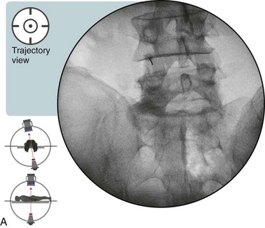
Tilt the fluoroscope cephalad or caudad, if needed.


 Trajectory View Safety Considerations
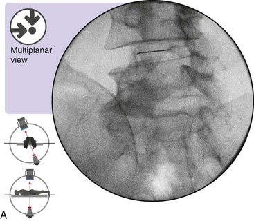
Place the needle parallel to the fluoroscopic beam.
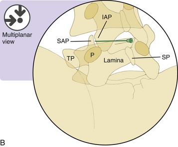
 Optimal Needle Position in Multiplanar Imaging
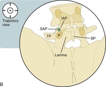
Optimal Needle Positioning in the Oblique View (Figure 13–2)
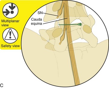
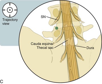
 Safety Considerations
Related posts:
 Atlantoaxial Joint Intraarticular Injection
 Lumbar Zygapophysial Joint Nerve (Medial Branch) Radiofrequency Neurotomy, Posterior Approach
Stay updated, free articles. Join our Telegram channel

Full access? Get Clinical Tree


