and Vincent L. Sorrell2
(1)
Division of Nuclear Medicine and Molecular Imaging Department of Radiology, University of Kentucky, Lexington, Kentucky, USA
(2)
Division of Cardiovascular Medicine Department of Internal Medicine Gill Heart Institute, University of Kentucky, Lexington, Kentucky, USA
Electronic supplementary material
The online version of this chapter (doi: 10.1007/978-3-319-25436-4_10) contains supplementary material, which is available to authorized users.
There are many causes of diffuse and focal “hot” and “cold” lungs on SPECT MPI (Table 10.1). Left ventricular dysfunction may be associated with diffusely increased lung uptake (Fig. 10.1) (Chamarthy and Travin 2010; Hendel et al. 1999; Mohr et al. 1996; Shih et al. 2002). Various benign pulmonary conditions, including infectious, occupational, and granulomatous processes (tuberculosis, sarcoidosis), may show diffusely or focally increased uptake of MPI radiopharmaceuticals (Aras et al. 2003; Chin et al. 1995; Gedik et al. 2007). Avidity may reflect disease activity in tuberculosis (Onsel et al. 1996).


Table 10.1
Differential diagnosis of “hot” and “cold” imaging findings related to the lungs
Organ system | “Hot” finding | “Cold” finding | References |
|---|---|---|---|
Lungs | Congestive heart failure (diffuse pattern) Interstitial lung disease Smoking Atelectasis Hematoma Pneumonia Fibrosing alveolitis Actinomycosis Granulomatous disease (sarcoidosis/tuberculosis) Neoplasm, primary malignant (lung carcinoma, carcinoid) Neoplasm, metastasis Lymphangitic carcinomatosis | Hyperinflation/emphysema Pneumonectomy/elevated right hemidiaphragm Elevated left hemidiaphragm | Aras et al. (2003) Bom et al. (1998) Chamarthy and Travin (2010) Chin et al. (1995) Eftekhari and Gholamrezanezhad (2006) Gedik et al. (2007) Hendel et al. (1999) Mohr et al. (1996) Nishiyama et al. (1997) Onsel et al. (1996) Reyhan et al. (2004) Seo et al. (2005) Shih et al. (2002) Takekawa et al. (1999) |
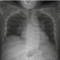
Fig. 10.1
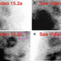
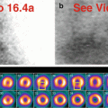
Congestive heart failure. An 86-year-old male with known coronary artery disease complains of shortness of breath and fatigue. MPI shows diffusely “hot” lungs (a–c), an enlarged right ventricular cavity and prominent right ventricular myocardial wall (a, b, d), a minimally enlarged left ventricular cavity (a, b, e), and depressed left ventricular function with global hypokinesis and LVEF of 40 % (f). The inferolateral-basal wall perfusion defect is more pronounced on rest images and normalizes with AC, suggesting diaphragmatic attenuation artifact (e). Cardiomegaly, a small right pleural effusion, and increased lung markings are evident on chest radiography (g). Echocardiography demonstrated concordant findings: left ventricular cavity enlargement, reduced function (LVEF of 30–40 %), and a slightly dilated right ventricle with reduced systolic function.(a) Rest raw projection images (Video 10.1a, frame 1), 99mTc sestamibi. (b) Stress raw projection images (Video 10.1b, frame 1), 99mTc sestamibi. (c) Stress raw projection image (Video 10.1c, frame 14), 99mTc sestamibi, lung activity (pink boxes). (d) Stress raw projection image (Video 10.1c, frame 43), 99mTc sestamibi, right ventricle (blue lines). (e) Stress/rest processed SPECT images (without and with AC) (SA, HLA, VLA), inferolateral-basal defect most pronounced on non-AC rest images normalizes on AC (yellow ovals on representative SA images, yellow boxes on representative HLA and VLA images). (f) Stress and rest gated SPECT images (Video 10.1d, frame 1) (SA, VLA, HLA). (g) PA chest radiograph
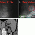
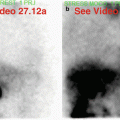
Through various mechanisms and local factors, primary and metastatic lung neoplasms accumulate MPI radiopharmaceuticals (Bom et al. 1998; Takekawa et al. 1999). Certain primary lung malignancies may be less avid for 99mTc sestamibi compared to 201Tl chloride (Nishiyama et al. 1997). Focal increased uptake in the lung may signal unknown lung cancer (Fig. 10.2) (Aras et al. 2003; Chin et al. 1995; Hendel et al. 1999; Shih et al. 2002). “Whole-field-of-view reconstruction” of the projection data set from Tc-99m sestamibi SPECT MPI has revealed a carcinoid tumor located behind the heart (Reyhan et al. 2004).


Fig. 10.2


Lung cancer. A 60-year-old female has a focal “hot” lesion in the posterior right lung (a–d) that corresponds to a discrete nodular mass on CT (e).(a) Stress raw projection images (Video 10.2a, frame 1), 99mTc tetrofosmin. (b) Stress raw projection image (Video 10.2b, frame 13), 99mTc tetrofosmin, lesion (pink oval). (c) Stress “whole-field-of-view” processed SPECT images, coronal from anterior to posterior, lesion (crosshairs in middle image). (d) Stress “whole-field-of-view” processed SPECT images, axial from superior to inferior, lesion (crosshairs in middle image), heart for reference (yellow box). (e) CT image through the lower lungs, lesion (pink box)
Related posts:

Stay updated, free articles. Join our Telegram channel
Full access? Get Clinical Tree
