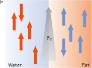
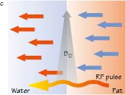
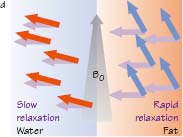
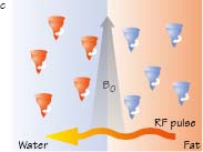
The body’s hydrogen nuclei behave as ‘bar magnets’ which are randomly aligned (a). When an external magnetic field ‘B0’ is applied to the body the magnetic vectors of most hydrogen nuclei line up along the field lines of B0 (b). When an RF pulse is then applied these vectors are tipped into a transverse plane (c). When this RF pulse is removed the vectors ‘relax’ to their initial longitudinal position (d). Detection of T1 MR signal occurs during this relaxation process and the final MR image comprises a graphic representation of differences in T1 characteristics of tissues. Hydrogen nuclei within tissues predominantly containing fat relax rapidly and produce high T1 signal, which is a measure of the return of hydrogen nuclei from transverse to longitudinal axis alignment. Fat therefore appears bright on T1-weighted images. Hydrogen nuclei within tissues predominantly containing water relax more slowly and produce a low T1 signal and therefore appear dark on T1-weighted images
5.2 T2 relaxation

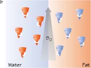
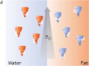
The body’s hydrogen nuclei are randomly aligned and are ‘spinning’ on their own axis (a). When an external magnetic field ‘B0’ is applied to the body (b) the nuclei align (as in Fig 5.1b) but also ‘precess’ (spin around their axis at a specific frequency related to the energy of B0). When an RF pulse is then applied (c) the spinning nuclei are forced into ‘phase’ (coherent synchronised spinning). When this RF pulse is removed however, the nuclei lose this phase coherence and ‘relax’ to return to their random phases (d). Detection of T2 MR signal tissue contrast occurs during this dephasing process and the final MR image comprises a graphic representation of differences in T2 characteristics of tissues. Hydrogen nuclei within tissues predominantly containing water dephase slowly and maintain high T2 signal. Water therefore appears bright on T2-weighted images. Hydrogen nuclei within tissues predominantly containing fat dephase more rapidly and therefore lose T2 signal. Fat is therefore less bright than water on T2-weighted images
Magnetic resonance physics
Related posts:


Stay updated, free articles. Join our Telegram channel

Full access? Get Clinical Tree


