Optimal treatment decisions for patients with rectal cancer are based on knowledge of tumor characteristics and prognostic features and any initial treatment must aim to reduce the risk of both local and distant recurrence. The radiologist has become an increasingly important part of multidisciplinary team managing rectal cancer. The primary goal of MRI staging of rectal tumors is to identify prognostic factors in order to offer patients a tailored treatment based on individual risks. Restaging of rectal tumors using MRI after chemoradiation therapy is becoming more relevant issue, since further tailoring of treatment is increasingly being considered after the treatment.
Key points
- •
High-resolution T2-weighted magnetic resonance imaging has emerged as the first-line imaging tool for multidisciplinary team decisions and is the most important sequence for evaluating rectal tumors.
- •
Rectal cancer staging is based on defining pertinent anatomy of the rectum and the surrounding structures, allowing for surgical planning and prognostic stage grouping.
- •
The primary goal of staging rectal cancer is to identify risk factors for distant or local recurrence to offer tailored treatments, based on individual prognosis.
- •
Some investigations are now focused on conservative management of rectal cancer, increasing the demand for radiologic evaluation of response to chemoradiation to distinguish responding from nonresponding tumors.
- •
Diffusion-weighted imaging may be a useful adjunctive tool for monitoring the response to chemoradiation therapy for rectal cancer.
Introduction and background
Cancer is a major public health problem in the United States and many other parts of the world. A total of 1,660,290 new cancer cases and 580,350 deaths from cancer are projected to occur in the United States in 2013. Among these cases, it is estimated that 102,480 will be new colorectal cases (39% from the rectum) and 50,830 will be cancer deaths. Worldwide, colorectal cancer is the third most common cancer in men (663,000 cases, 10.0% of the total) and the second in women (571,000 cases, 9.4% of the total). Recent trends in the United States show a decreasing incidence of invasive colon and rectal cancers in both men and women, which can be attributed to adoption of colorectal screening programs with earlier detection and removal of precancerous polyps.
Historically, prognosis of rectal cancer has been directly related to the extent of extramural spread into the mesorectum and the ability to achieve surgical clearance at the circumferential resection margins (CRMs), and for patients not undergoing total mesorectal excision (TME) surgery, pelvic recurrence rates were strongly linked to nodal status. Quirke and colleagues reported that microscopically positive resection margins occurred in up to 40% of patients treated by non-TME surgery, with local recurrence rates of 83%. Two advances in therapy have a substantial effect on reducing the frequency of local recurrence and improving survival: TME and preoperative neoadjuvant chemoradiation therapy (CRT). The adoption of preoperative radiation therapy for advanced tumors showed a substantial reduction in pelvic recurrence rates of clinically resectable rectal cancers from 20% to 10% in the first Stockholm trials, and this was associated with a survival benefit. More recently, the widespread adoption of TME techniques has reduced the rates of margin involvement in unselected rectal cancers undergoing TME surgery from up to 28% to less than 15% in the last decade. Therefore, precise assessment of the distance of tumor to the mesorectal fascia by preoperative staging has become important for distinguishing between patients who will be cured by primary surgery and those who are at high risk for local disease recurrence because of the risk of circumferential margin involvement.
High-resolution magnetic resonance (MR) imaging plays a pivotal role in the pretreatment assessment of the most important risk factors for local recurrence. However, another important role of MR imaging is in the evaluation of low rectal tumors, in which sphincter preservation is a challenge. Assessment of the safety of the TME plane is crucial, because sphincter function may be severely compromised by irradiation, but, on the other hand, perforation of a low rectal cancer during the TME dissection results in local recurrence that could be avoided by preoperative shrinkage of the tumor through radiotherapy. Because the mesorectum significantly tapers toward the top of the anal canal, tumors in this area can easily invade surrounding structures. Therefore careful assessment of the safety of the TME plane by MR imaging is essential. In a selected group of patients, CRT with delayed surgery increases the likelihood of preserving sphincter function, because of a downsizing and downstaging effect of induction therapy on the tumor, leading to improved resectability and local control. Tumor shrinkage as a result of preoperative CRT is now a reality, and pathologically complete responses (CRs) are not uncommon.
The success of this imaging technique depends on obtaining good-quality high-resolution T2-weighted images of the primary tumor, the mesorectal fascia, and mesorectal and pelvic sidewall lymph nodes. MR imaging can predict the CRM with high accuracy and consistency, allowing preoperative identification of patients at risk of recurrence that benefits from preoperative treatment, more extensive surgery, or both. The technique also enables the identification of patients whose disease is optimally treated with primary total mesorectal surgery alone, with preservation of the sphincter complex. Recent studies have shown that it is a reliable and reproducible technique with high specificity (92%) for predicting a negative CRM, the relationship of the tumor to the CRM, and the depth of tumor invasion outside the muscularis propria. Furthermore, the MERCURY (Magnetic Resonance Imaging and Rectal Cancer European Equivalence) trial centers were able to select up to 33% of patients with good prognostic features who could undergo primary surgery without preoperative therapy and without developing local recurrence on long-term follow-up.
There are data indicating that pathologic response after preoperative chemoradiotherapy is a prognostic factor for disease-free survival (DFS) for advanced rectal cancer and magnetic resonance is a valuable tool to evaluate the tumor regression grade (TRG). Moreover, there are some observational data that indicate that in patients with clinical CR, surgery may be avoided. Careful follow-up with a wait-and-see approach has produced impressive results, similar to those of radiation therapy for anal carcinoma. These studies have used biopsy or excision of the scar to define CR. The potential role of MR imaging in not only identifying patients who are likely to have CR but also in monitoring those patients for tumor regrowth is under investigation in a prospective clinical trial (Deferral of Surgery trial, UKCRN ID 8565, National Cancer Research Institute, UK).
So, with the increasing availability of preoperative therapy and the proven ability of MR imaging to give accurate prognostic information, the radiologists’ role in the preoperative multidisciplinary team decision-making process has become critical, because the information provided by the detailed imaging of the primary tumor guides the team to help achieve better outcomes for patients with rectal cancer.
Normal anatomy
Over the past years, the surgical anatomy of the pelvis has been reevaluated, because there is growing evidence of the benefits related to careful anatomic dissection in rectal cancer surgery. The complete removal of the tumor-containing rectum and its draining nodes as a distinct anatomic package is the essence of TME and has resulted in reduced local recurrence rates.
The rectum is that part of the gastrointestinal tract that extends from the upper end of the anal canal to the rectosigmoid junction and is approximately 15 cm in length. Anatomically, it can be divided into 3 segments: the low, mid, and high rectum. These segments correspond to the first 7 to 10 cm, the next 4 to 5 cm, and the last 4 to 5 cm (measuring from the anal verge), respectively.
The proximal part of the anal canal is characterized by the insertion of the levator ani muscle onto the fibers that form the puborectalis sling. Recognition of the inferior limits of the rectum is important in determining the distance between the tumor and the puborectalis sling, which is crucial for sphincter preservation during surgery. There are some important structures that must be recognized on MR imaging scans: the rectal wall layers, the mesorectum, the mesorectal fascia, the retrorectal space, the rectosacral fascia, the peritoneal reflection, and the Denonvilliers fascia.
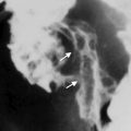
In cross section, the rectal wall consists of the mucosal layer, muscularis mucosae, submucosa, and muscularis propria ( Fig. 1 A). Most of the rectum lies below the peritoneal reflection, and so only the upper third is invested by the serosa or peritoneum. On MR imaging, the mucosal layer of the bowel wall is visible as a fine, low-signal-intensity line with the thicker, higher-signal-intensity submucosal layer beneath. The muscularis propria is shown on MR imaging as 2 distinct layers: the inner circular and the outer longitudinal layers. The outer layer has an irregular appearance and some surface interruptions caused by vessels entering the rectal wall, but readily distinguished from tumor by its lower signal intensity on high-resolution T2-weighted images.

The mesorectum is the fatty tissue shown on axial MR imaging as a high-signal-intensity structure that surrounds the rectum and contains lymph nodes, lymphatics, and vessels and is encircled by the mesorectal fascia, which represents the CRM when TME is used as the surgical technique. The mesorectal fascia is a layer encompassing the mesorectum, and it is best seen on axial and coronal MR images, appearing as a low-signal-intensity thin linear structure. It is optimally visualized on high-resolution thin-section MR imaging (see Fig. 1 B). Benign lymph nodes within the mesorectum are shown as uniform-signal-intensity ovoid structures, with smooth-bordered margins and homogeneous texture. Benign or reactive nodes are predominantly high signal intensity on the T2 sequences, but can also be uniformly low or intermediate signal intensity and can be significantly enlarged despite being benign. Sometimes, a regular chemical shift artifact can be seen in 1 side of it.
The parietal fascia fuses with the sacral periosteum at the level of the sacral promontory and within the pelvis overlies the muscles of the pelvic wall. Anteriorly, this fascia is attached to the body of the pubis. On MR imaging, it appears isointense relative to the muscles and may not always be seen as a separate structure, but it can be clearly seen as a separate layer overlying the pelvic sidewall compartment and between the mesorectal fascia and presacral compartment ( Fig. 2 ).

The retrorectal space is situated between the presacral parietal fascia and the mesorectal fascia. The presacral fascia is shown on sagittal MR imaging as a low-signal-intensity thin linear structure overlying the presacral vessels ( Fig. 3 ). The mesorectal fascia is seen anterior to this structure, and the potential virtual space between these 2 fascial layers forms the retrorectal space and the plane of dissection in TME surgery.

The rectosacral fascia is a pelvic floor fascial structure of variable thickness, which is shown on sagittal MR imaging as an oblique low-signal-intensity band extending from the junction of the S3 and S4 vertebrae to the posterior wall of the rectum, adjacent to the anal sphincteric complex ( Fig. 4 ).

The peritoneal reflection is easily seen on sagittal MR imaging as a low-signal-intensity thin linear structure that extends over the surface of the bladder posteriorly to its point of attachment onto the rectum. In men, the attachment site of the peritoneal reflection is the junction of the upper two-thirds and lower one-third of the rectum (see Fig. 4 ). In women, this site of attachment has more anatomic variations. The relationship between the rectal tumor and the peritoneal reflection is important in staging of the rectal cancer, because the tumors with invasion through the peritoneal reflection are categorized as stage T4a.
The Denonvilliers fascia is a well-developed fascia that derives from the urogenital septum during embryonal development. It forms a characteristic anterior surface of the mesorectum, on its lower part, and it is visible on axial MR imaging as a low-signal-intensity layer, adjacent to the prostate in men and as the rectovaginal septum behind the posterior vaginal wall in women ( Fig. 5 ). Inferiorly, the septum extends to the perineal body.

Imaging technique
Before the development of the modern phased-array pelvic surface coil, endorectal coils were the only method of obtaining high-resolution images of pelvic anatomic structures. The use of endorectal coils was limited in rectal cancer assessment, because of near-field artifact and luminal distortions created by the coils in direct contact with the rectal wall. Further problems with endorectal coils, as with any endoluminal techniques such as endoanal ultrasonography, were the inability to evaluate stenosing tumors and to fully evaluate the entire mesorectal lymph node drainage territory.
The introduction of phased-array coil systems improved staging of rectal cancer, which along with fast spin-echo T2-weighted sequences enabled high-resolution imaging oriented to the relevant surgical anatomic planes. These phased-array surface coils combined with a very high spatial resolution allowed detailed evaluation of the rectal wall and depiction of the surrounding important anatomy.
The technique for acquisition of the sequences has been previously described, and the parameters are shown in Table 1 .
| Sequence | FOV (mm) | Repetition Time/Echo Time | Slice (mm) | Matrix | NSA | Time (min:s) | |
|---|---|---|---|---|---|---|---|
| Sagittal T2 From one pelvic sidewall to the other | TSE/23 | 250 | 3500/125 | 3 interleaved | 304 × 512 | 4 | 6:36 |
| Axial T2 Iliac crest to pelvic floor | TSE/22 | 420 | 3500/80 | 5 interleaved | 352 × 512 | 1 | 1:38 SENSE 2 |
| Axial T2 Perpendicular to the rectum Oblique axial T2 To cover lymph node territory | TSE/16 | 160 | 3500/120 | 3 interleaved | 256 × 512 | 6 | 5:36 |
| Coronal T2 Low tumors parallel to anal canal | TSE/16 | 160 | 3500/120 | 3 interleaved | 256 × 512 | 6 | 5:36 |
A 1.5-T system is generally used with phased-array coils, which allow greater coverage of the anatomy when compared with the endorectal coils. The experience with a 3-T system in the high-resolution protocol is still limited, but it is likely that there are few or no benefits with this system for the staging of rectal cancer.
Patients must be fully informed about the length of time required for MR imaging scanning (between 20 and 30 minutes) and must be positioned in the supine position in the scanner. There is no need to use purgative bowel preparation or enemas. Antispasmodic agents are used to reduce pelvic small bowel motion. Gadolinium-enhanced T1-weighted imaging and fat saturation are not necessary, because these have not been shown to improve the diagnostic accuracy for staging of rectal cancer.
Initial localization images in the sagittal planes are needed to plan the high-resolution images ( Fig. 6 A–C). For this reason, the first series to be acquired is a sagittal, 25-cm field-of-view (FOV), 3-mm-thick T2-weighted sequence from 1 pelvic sidewall to the other, which enables identification of the primary tumor. It is essential that the referring surgeon has accurately indicated the tumor position (low, mid, or high rectal) to help proper planning of the sequences. The second series consists of large FOV axial sections (4–5 mm) of the whole pelvis. These first 2 sequences allow an overview of the pelvis and enable the primary tumor to be located with regard to the distance from the anal verge and puborectalis sling, but these are not adequate for T staging or nodal characterization, which are best undertaken using the high-resolution sequences described later.

The high-spatial-resolution sequence comprises a T2-weighted thin axial section (3 mm) through the rectal tumor and adjacent tissues. These sequences are angled perpendicular to the long axis of the rectum at the level of the tumor, using a 16-cm FOV (see Fig. 6 B, D). The perpendicular plane is necessary; otherwise, the images may be misinterpreted because of partial volume effects. This sequence must be extended to at least 5 cm above the superior edge of the tumor to ensure coverage of draining nodes and tumor deposits (see Fig. 6 A, C). Knowledge of the distribution pattern of mesorectal nodes may assist in preoperative radiotherapy planning if the tumor shows high-risk features.
The T2-weighted thin-section coronal sequence is optional for the upper-rectum and midrectum tumor, but mandatory for patients with low rectal cancers. This sequence consists of high-spatial-resolution thin sections (3 mm) parallel to the long axis of the rectum at the level of the tumor and it shows the levator muscles, including the puborectal, and the sphincter complex in relationship to the rectal wall ( Fig. 7 ).

The routine use of diffusion-weighted imaging (DWI) is gradually increasing and preliminary data suggest that apparent diffusion coefficient (ADC) values may reflect tumor aggressiveness, but it is not clear whether this provides additional prognostic information compared with conventional T2-weighted high-resolution imaging and whether ADC values could influence treatment decisions. The published data suggest that it is unlikely that the addition of DWI alters staging accuracy because of substantial overlap in ADC values between benign and malignant processes.
Water diffusion is a physical process that results from the thermally driven, random motion of water molecules. In a glass of water, molecules undergo free, thermally agitated diffusion (with a three-dimensional Gaussian distribution). The width of the Gaussian distribution expands with time, and the average square of this width per unit time gives the units of the ADC. In tissues, apparent diffusion is observed because the movement of water molecules is modified by their interactions with cell membranes and macromolecules in an environment. The restriction to flow of water molecules is determined by tissue cellularity and the integrity of the cell membrane. Tumors typically display restricted diffusion because of their hypercellularity.
In our standard 1.5-T GE Signa HDxt system (General Electric Medical Systems, Milwaukee, WI), we use an 8-channel cardiac coil, with a spin-echo echo planar imaging sequence (repetition time [TR], 4500 milliseconds; echo time [TE], 80 milliseconds; flip angle, 90°; FOV, 280–360 mm; 16 number of averages; slice thickness, 4 mm; interslice gap, 0.4 mm; acquisition matrix, 128 × 100) with bandwidth of 1.953 Hz/pixel, with an imaging time of DWI of 3 minutes 36 seconds for 20 slices, b value (0, 500, 1000 s/mm 2 ), and 3 directions. DWI is performed in the axial plane. The motion-probing gradient pulses are placed in the x-axis, y-axis, and z-axis (all planes). The parameters are shown in Table 2 .
| Parameter | Philips | Siemens | General Electric |
|---|---|---|---|
| FOV (cm) | 280 | 220 | 280 |
| Matrix size | 112 × 256 | 138 × 256 | 128 × 100 |
| TR | 2500 | 3100 | 4500 |
| TE | 66 | 71 | 80 |
| Parallel imaging factor | 2 | 2 | 2 |
| Number of signals average | 4 | 8 | 16 |
| Section thickness (mm) | 5 | 5 | 4 |
| Direction of motion-probing gradients | 3 scan trace | 3 scan trace | 3 scan trace |
| Fat suppression | SPAIR | SPAIR | STIR |
| b factors (s/mm 2 ) | 0,350,750 | 0,350,750 | 0,500, 1000 |
Imaging interpretation
Staging of the Primary Rectal Tumor
The American Joint Committee on Cancer guidelines, seventh edition, defines the criteria for the staging of primary rectal tumors. The process of T staging, T substaging, and N staging is shown in Table 3 . The T staging is based on the invasion of the primary tumor through the rectal wall and its relationship to the submucosa and muscularis propria. Therefore, the best results for staging rectal cancer have been obtained through careful interpretation of thin-section, high-resolution, and small FOV T2-weighted images obtained perpendicular to the rectal wall.
Related posts:

Stay updated, free articles. Join our Telegram channel

Full access? Get Clinical Tree


