Fig. 1
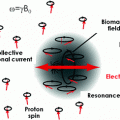
Light from a circularly polarized laser beam optically pumps the Rb atoms, while light from a second laser beam probes the magnetic field-dependent spin orientation through polarization rotation. Both the transmission and polarimeter signal show a resonance as a function of magnetic field
For MEG measurements made thus far, only zero-field magnetometers have been used. When operated in a regime of frequent atomic collisions and in low magnetic fields, the decoherence mechanism through spin-exchange collisions can be suppressed (Happer and Tang 1973). This so-called spin-exchange relaxation-free (SERF) regime (Allred et al. 2002) allows for very high magnetometer sensitivities, but limits the dynamic range of the magnetometer. In very small magnetic fields, the spins are tilted by the magnetic field and a static reorientation results from the balance between precession and continuous pumping. Again, the orientation of the spins is measured with near-resonant light by detecting the transmission of resonant light. Often it is more advantageous to monitor the polarization rotation of a slightly detuned light beam, which is usually done with a balanced polarimeter. This method can cancel intensity noise of the laser light and also tolerate much higher optical thickness of the vapor, since the light is detuned from resonance. In order to increase the signal-to-noise ratio, phase-sensitive detection can be implemented. For the zero-field resonances, one parameter, such as the magnetic field or the probe light polarization, is modulated and the same frequency component of the light is detected at a fixed phase with the modulation. Typically, OPMs measure the magnitude of the magnetic field, but the zero-field OPMs used for MEG are operated to measure a magnetic-field component in a certain direction, e.g., through external field modulation with an additional Helmholtz coil pair.
All of the devices used for MEG demonstrations have been operated in an open-loop configuration, where the dynamic range was limited by the linewidth of the resonance to below 100 nT. While feedback systems have been demonstrated in principle, they have not yet reached the same sensitivities (Seltzer and Romalis 2004). Furthermore, the response of the magnetometer shows the behavior of a first-order low-pass filter with a width corresponding to the linewidth of the atomic resonance. This usually limits the intrinsic bandwidth to below 1 kHz.
Figure 2a shows a chip-scale high-sensitivity OPM, which was manufactured by use of a micro-electro-mechanical-system (MEMS) process (Mhaskar et al. 2012). In the center of the cube is an alkali-vapor cell, heated to generate a sufficient atomic density of the vapor (Knappe et al. 2005). A laser, on resonance with a transition of the atoms, is circularly polarized and optically pumps the atoms. The same laser is used to monitor the atomic polarization by detecting the transmitted light with a photodiode (Dupont-Roc et al. 1969; Shah et al. 2007). This is only one of many possible configurations with respect to polarization, sensor shape, and beam.
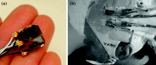

Fig. 2
a Photograph of a chip-scale OPM sensor head. b Photograph of four chip-scale OPMs attached to an EEG cap for MEG measurements. The fibers and wires needed to drive a sensor are collected in a bundle leaving the head tangentially
3 MEG with OPMs
Three different demonstrations of MEG measurements with OPMs on human subjects have been published to this date. In the first one, Xia et al. used a (7.5 cm)3 Pyrex cell filled with potassium vapor and heated to 180° C (Xia et al. 2006). It was placed on the left side of the head at a distance of 6.25 cm between the scalp and center of the cell. The atoms were polarized with 500 mW circularly polarized pump light, and the magnetic field was monitored through the polarization rotation of linearly polarized probe light at a right angle, so that the OPM was sensitive to the component of the magnetic field normal to the scalp. The probe light was detected by a 16 × 16 photodiode array and 256 parallel channels of roughly 0.4 × 0.4 × 7.5 cm and spacing of 5 mm could be monitored simultaneously. Auditory evoked fields were recorded and the N100 m peak was clearly visible in the data. Six subjects were measured.
In the second paper Johnson et al. demonstrated an OPM with a fiber-coupled sensor head. Pump and probe beams were collinear and passed through a cylindrical Rb vapor cell of diameter 2.5 cm and length 2.5 cm (Johnson et al. 2010). A balanced polarimeter consisted of a photodiode and a quadrant detector, which allowed them to monitor four channels with a spatial distance of 5 mm simultaneously. The distance between cell and scalp was 2 cm, and one component of the magnetic field tangential to the scalp was measured. Evoked fields were recorded after auditory stimulation and after electrical stimulation of the median nerve in a male subject. They were verified by consecutive SQUID measurements.
Stay updated, free articles. Join our Telegram channel

Full access? Get Clinical Tree


