Gender (M/F)
47/59
Age (year), range (median)
26–79 (63)
GTV (mL), range (median)
1.0–325.8 (28.3)
Tumor site
Oral
12
Nasal
68
Paranasal sinus
18
Pharynx
5
Conjunctiva
3
Tumor status (UICC2002a)
T1
9
T2
18
T3
30
T4 (a, b)
34
Recurrence after surgery/chemotherapy
15
Table 16.2
DAV chemotherapy
Number of chemotherapy courses | |
5 courses completed | 57 |
1–4 courses | 49 |
Reasons for discontinuing chemotherapy | |
Recurrence and/or metastasis | 16 |
Side effects | 33 |
16.5.2 Toxicity
No severe acute reactions were observed. Acute reactions of the normal tissue in the 106 patients included grade 3 skin reactions in one patient (0.9 %) and grade 3 mucosal reactions in 23 patients (21.7 %); no reactions worse than grade 3 were observed.
Late toxic reactions were grade 2 mucosal reactions in two patients (1.9 %), with no evidence of toxicities worse than grade 2.
The grades and incidence rates of radiation-induced side effects were similar to those in patients with head and neck tumors (previous chapter) and did not seem to be exacerbated by DAV chemotherapy.
The most common adverse events from DAV chemotherapy were myelotoxicity, liver dysfunction, transient nausea, vascular pain, and photosensitivity.
Of the 106 treated patients, 15 patients (14.2 %) had grade 3 myelotoxicity events and four patients (3.8 %) had grade 3 or 4 liver dysfunction events. The patient with grade 4 liver dysfunction was found shortly thereafter to have multiple hepatic metastases. Thus, the patient’s liver function might have been affected by the metastases.
No treatment-related deaths occurred during the study.
16.5.3 Local Control and Overall Survival
The median survival time was 64.8 months. The 3- and 5-year overall survival rates for all patients were 65.8 and 54.0 %, respectively, and the 3- and 5-year local control rates for all patients were 83.1 and 80.6 %, respectively.
Since 1997, C-ion RT has been used for MMM of the head and neck. After C-ion RT, many patients developed distant metastasis, although local recurrence was not noted [25, 26]. This finding indicates that, in these patients, systemic micrometastases might have already existed at the time of treatment, and management of these events would be desirable for improved survival. On the basis of this result, we have begun to administer C-ion RT concomitantly with DAV chemotherapy. Consequently, although no difference was observed in the local control rate, the survival rate was significantly improved by DAV chemotherapy. C-ion RT with concomitant DAV chemotherapy is a safe and effective treatment for MMM of the head and neck because of good local control and acceptable toxicity.
16.6 Case Study
16.6.1 Case 1
A 61-year-old male patient presented with a growth in the palatal region and reported pain for a 2-month duration. The patient was admitted to the Oral and Maxillofacial Surgery outpatient clinic. Biopsy indicated a diagnosis of malignant melanoma. Melanosis was observed mainly in the left hard palate.
Tumors without thickness cannot be measured by MRI and CT. We devised a method with which to clarify the presence of the tumor by CT image fusion with a tumor outline that had been marked with metal wire (Fig. 16.1a, b).
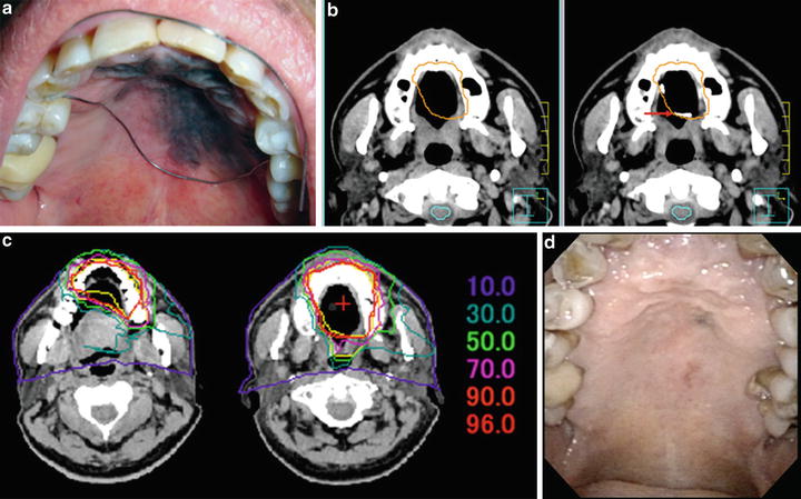
Fig. 16.1
Left palate MMM in a 61-year-old male patient. (a) Intraoral view of the lesion and placement of a metal wire (arrow) to clearly outline the tumor. (b) Image fusion of the planning CT (left) and CT with the wire (right). (c) Dose distribution of C-ion RT. (d) Intraoral view 2 years after C-ion RT
The patient received C-ion RT and DAV chemotherapy. A total radiation dose of 57.6 GyE in 16 equal fractions was administered over 4 weeks (Fig. 16.1c). Acute mucositis was reduced by reducing the dose to the dorsum of the tongue.
DAV chemotherapy was administered in five courses. Grade 2 leukopenia and grade 1 hepatic dysfunction were transiently experienced. However, these events were quickly resolved, with level returning to within the normal range.
The patient was followed up for 2 years after C-ion RT. Local recurrence and distant metastasis were not observed (Fig. 16.1d).
The presence of teeth in the irradiation range increases the risk of osteonecrosis. It is important that oral hygiene guidance be provided with the assistance of a dentist and dental hygienist.
16.6.2 Case 2
A 66-year-old female patient consulted the Department of Otolaryngology because of a left nasal obstruction and epistaxis. Physical examination revealed a measurable black tumor in the left nasal cavity. Imaging modalities also showed a tumor in the left nasal cavity. Biopsy indicated a diagnosis of malignant melanoma (Fig. 16.1a, b). The patient consulted our hospital in order to undergo C-ion RT.
The patient received C-ion radiotherapy and DAV chemotherapy. A total radiation dose of 57.6 GyE in 16 equal fractions was given over 4 weeks (Fig. 16.2c


Related posts:

Stay updated, free articles. Join our Telegram channel
Full access? Get Clinical Tree
