Management of Trauma to the Liver and Spleen


Management of Trauma to the Liver and Spleen


Robert F. Dondelinger


Clinical examination is unreliable in both establishing the diagnosis of hepatic or splenic contusion and evaluating its severity. In the most severe injuries, during the initial phase of compensated hemorrhagic shock, clinical signs are subtle until 40% of the blood volume is lost. The ensuing bradycardia can be puzzling in that it may be observed as a result of vagal stimulation. Because of their rich vascularization, both the liver and spleen can bleed profusely and be responsible for rapidly progressive hypotension or shock. If blood volume is not corrected quickly, uncompensated hemorrhagic shock occurs, followed by metabolic acidosis, myocardial depression, diffuse intravascular coagulation, and death. Polytransfusion and hypothermia further contribute to the metabolic acidosis and wash-out coagulopathy that can jeopardize the result of arterial embolization.


In patients with less severe hepatic or splenic injury, abdominal pain, either diffuse or localized to the right or left upper quadrant, is the most frequent complaint. Localized abdominal symptoms are found in 27% and nonspecific abdominal symptoms in 41%. Pain in the right shoulder (Kehr sign) or its equivalent on the left side is indicative of phrenic, hepatic, or biliary injury on the right and splenic injury on the left and is specific if no trauma to the shoulder has occurred. The presence of three or more lower rib fractures increases the relative risk for hepatic or splenic injury significantly and is an indicator for thorough imaging. Respiratory difficulty caused by limited diaphragmatic excursion due to pain or phrenic irritation is a symptom suggestive of underlying liver or spleen injury. Occasionally, hemothorax is a sign of splenic rupture because a lacerated spleen is likely to herniate through a phrenic tear and bleed into the thorax. It should be stressed that clinical symptoms can be absent in up to 19% of patients with documented liver injury and in 35% with splenic contusion. Elevation of serum transaminase levels in excess of 130 IU/L is an indication of liver injury, even in the absence of clinical complaints. Posttraumatic hemobilia and bilhemia are of particular interest for the management of patients with liver injury.


Hemobilia is a sign of intrahepatic hemobiliary fistula and is estimated to develop in 2.5% of patients with hepatic injuries. Posttraumatic hemobilia is generally caused by an intrahepatic vascular lesion, such as a pseudoaneurysm or arteriovenous fistula (AVF) that has eroded a bile duct wall. Therefore, hemobilia often coexists with biloma or a bile fistula. In some patients, persistent discrete extrahepatic hemorrhage is responsible for development of hemorrhagic ascites some time after the trauma. Persistent mixing of bile with blood from a liver contusion prevents healing and explains recurrences of hemobilia. Significant bile duct obstruction due to clot and causing jaundice can occur but is a rare event. Bleeding is quite variable: profuse, causing shock and requiring urgent treatment, or moderate or occult. With persistence of the lesion, which often goes unrecognized, hemobilia has a tendency to recur, sometimes for decades. The mean reported interval between injury and hemobilia is about 1 month, but a considerable time interval may separate the injury from onset of symptoms. The classic clinical triad consists of gastrointestinal hemorrhage, obstructive jaundice, and biliary colic. The triad is incomplete in about 60% to 70% of cases.


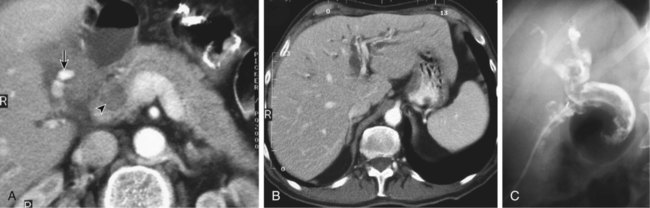
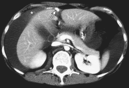
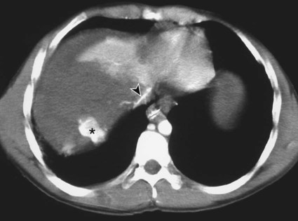
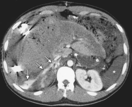
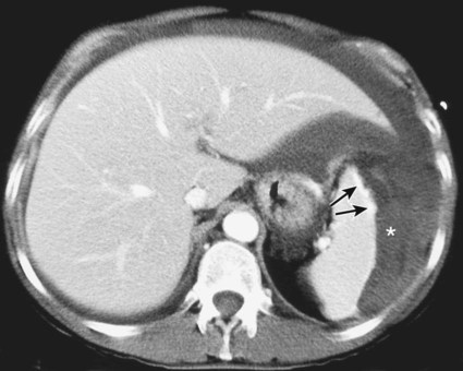
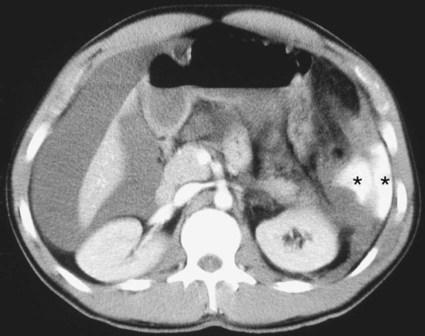
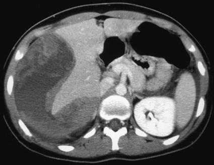
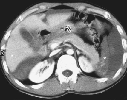
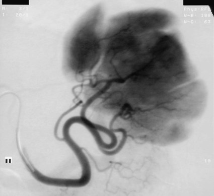
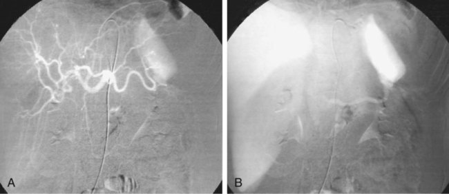
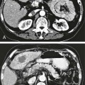
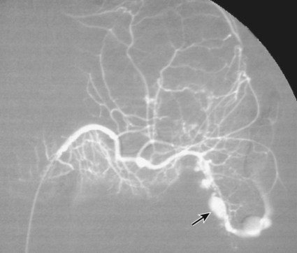
The diagnosis of hemobilia is indirectly established by endoscopy. Endoscopic retrograde cholangiopancreatography (ERCP) confirms bleeding from the papilla in only a minority of cases, but it shows intraluminal choledochal defects corresponding to clots, also occasionally seen in the pancreatic ducts, and provides evidence of proximal biliary obstruction (Fig. 71-1). ERCP does not show the intrahepatic bleeding site or the nature of the vascular lesion responsible for the hemobilia. Overall, ERCP is diagnostic of hemobilia in about a third of cases and can rule out a gastroduodenal bleeding source. Bile duct dilation often remains moderate because the clots are not totally obstructive. Endoscopic ultrasound (US) may be more contributory than ERCP by more precisely showing mobile hyperechoic material without acoustic shadows in the gallbladder and common bile duct, indicative of an endoluminal clot. Sphincterotomy with choledochal flushing removes clot from the bile ducts and exposes fragmented clot to the fibrinolytic activity of bile, thereby accelerating regression of the bile duct obstruction. Most often, the intrinsic fibrinolytic properties of bile cause clots to dissolve spontaneously. US, computed tomography (CT), and magnetic resonance imaging (MRI) can also suggest the diagnosis of hemobilia by showing echoic material in the bile ducts or hyperdense material and extra- and intrahepatic bile duct dilation and sometimes the intrahepatic vascular lesion. Blood pool scintigraphy may show accumulation at the site of an intrahepatic vascular lesion, but arteriography is definitive for both diagnosis and treatment. It locates the bleeding source in more than 90% of cases by demonstrating either an underlying vascular lesion—a pseudoaneurysm or an arterioportal fistula—or, in a minority of cases, extravasation of contrast material into the bile ducts. Spontaneous definite resolution of hemobilia without any type of intervention occurs in only a minority of patients.


Bilhemia results from a posttraumatic communication established between a bile radicle and an intrahepatic vein and is a rare event after blunt hepatobiliary injury, with only a limited cohort of patients being reported. Because of low pressure in the hepatic venous system, bile drains into the vascular system. Jaundice caused by bilhemia cannot always be distinguished from that caused by hemobilia. It may well happen that a patient has a massive increase in direct bilirubin early after trauma, as well as later in the course of follow-up for hemobilia, thus indicating that bilhemia and hemobilia may occur in the same patient. In contrast to hemobilia, in bilhemia ERCP is able to show passage of bile in hepatic veins, and hepatic arteriography is usually nondiagnostic.


Surgical treatment has shown that simple débridement and tamponade of the biliovenous fistula is therapeutic, and hepatic resection can be avoided. Bilhemia can be treated nonsurgically by selective endoscopic intrabiliary balloon placement to tamponade the leak within the liver for several days, or by sphincterotomy and bile duct stenting for decompression. If biliary obstruction is present and responsible for elevated pressure in the bile ducts, balloon dilation or stenting of a stricture should be performed.


Indications


Indications for hemostatic arterial embolization of hepatic or splenic posttraumatic injuries are not based on scientific evidence but rather on pragmatic grounds according to local logistics. Isolated hepatic and splenic injuries are most often considered in adults and in children.


In a hemodynamically stable or rapidly stabilized patient after resuscitation, the observation on CT of dense extravasation of contrast material or a localized blush of contrast material within the liver or spleen parenchyma or diffusing through the capsule and diluting in the hemoperitoneum in the subphrenic, perihepatic, or perisplenic space is an unequivocal sign of ongoing bleeding or free intraperitoneal hemorrhage (Figs. 71-2 to 71-6). Inside the liver or spleen parenchyma, contrast material may line the capsule or layer in a hematoma. Extravasated contrast material shows characteristic attenuation values of 80 to 130 Hounsfield units (HU) and might be surrounded by hematoma with lower density. The density of extravasated contrast material is similar to that measured in large opacified vessels. Clotted blood, in the absence of extravasation, has density values around 60 HU, and unclotted free intraperitoneal blood, not mixed with contrast material, has values of 35 to 45 HU (Fig. 71-7). Extravasation of contrast material usually appears rapidly at the arterial phase of opacification. Massive and rapid accumulation of contrast material in the perihepatic or perisplenic spaces without continuity from an intravisceral contusion indicates rupture of a hilar vessel. Rarely, extravasation appears only on delayed scans.


Indications for hepatic or splenic arteriography and embolization can be extended to patients with evidence of continuous hemorrhage who remain borderline after resuscitation. Today, because of rapid scanning by multidetector CT, all trauma patients suspected of sustaining abdominal injury undergo CT examination at admission. Such management requires optimal organization and skills in the radiology department. Patients with early ongoing bleeding after primary surgical hemostasis and those who are referred after crash laparotomy should also undergo arteriography without delay, being transferred immediately from the operating room to the angiography suite. Patients who rebleed after initially successful embolization should be treated again angiographically. After the initial posttraumatic phase, cross-sectional imaging is also able to demonstrate mature nonacutely bleeding vascular lesions such as pseudoaneurysms or AVFs in the liver or spleen. Because of their relatively high risk of delayed bleeding, these vascular injuries are likewise treated by selective embolization during a scheduled procedure.


Contraindications


A contraindication to hemostatic arterial embolization of the liver or spleen is a hemodynamically unstable patient who requires urgent regular or damage-control laparotomy.


There might be several pitfalls in recognizing extravasation of contrast material on CT. In the case of severe hypovolemic shock, the arteries contract as a result of the output of vasoconstrictive agents. Severe arterial vasoconstriction is called hypoperfusion complex on CT images and may simulate disruption of the splenic artery by decreased enhancement or complete absence of splenic enhancement (Fig. 71-8; also see Fig. 71-4). This syndrome occurs in 0.01% to 2% of cases of major abdominal trauma in children with hypovolemic shock and may lead to unnecessary laparotomy. The CT appearance is particularly puzzling when no splenic enhancement at all is seen, but other solid organs are normally enhanced. The liver with its dual vascular supply is not usually affected, but the pancreas can be affected as an isolated organ in the same way as the spleen. When vasoconstriction resolves, progressive splenic enlargement may be seen on repeat CT examination. A sudden increase in intrasplenic blood inflow in an injured vessel may be responsible for early rebleeding despite apparent initial hemostasis.


Theoretic causes of erroneous interpretation of hepatic or splenic injury on angiography include a false-positive diagnosis suggested by a splenic fissure; a nonopacified splenic polar artery; early venous return in the spleen caused by massive injection of contrast medium; superimposition of the gastric fundus, pancreatic tail, accessory spleen, or left adrenal; and preexisting hepatic or splenic infarct (Fig. 71-9). Because of the large projection area of the liver, false-positive results on arteriography are rare. False-negative diagnoses are due mainly to minimal parenchymal injury. False-negative findings of persistent hemorrhage are estimated to occur in 1% to 2% of patients. A multicenter survey found that arteriography is used today as a diagnostic modality in only 2% of patients being treated by observation.


Technique


Anatomy and Approach


Hepatic arterial anatomy and variants must be kept in mind to obtain a complete arteriographic workup. As a reminder, the right hepatic artery, right lateral hepatic artery, or celiac artery may originate from the superior mesenteric artery, and the left hepatic artery or branches of the left lateral sector may originate from the left gastric artery. Vascular anatomy and variants of the liver should be known to correctly diagnose any parenchymal filling defect due to a nonopacified aberrant hepatic artery. The origin of the cystic artery should be recognized because inadvertent embolization should be avoided. Rarely, bleeding from the cystic artery requires hemostatic embolization.


The spleen is usually vascularized through a single splenic artery that branches at the hilum. A polar artery, most often directed to the upper pole, may originate from the main artery proximal to the hilar branches. An accessory spleen has its own independent small artery originating from the main splenic artery at the hilum. Occasionally the hepatic and splenic arteries may have a separate ostium originating from the abdominal aorta.


Angiographic access is gained by either a femoral or left axillary approach.


Technical Aspects


Arteriographic Findings


Arteriography was used to evaluate hepatic or splenic injury in the past, but rapidly became obsolete with the advent of US and CT. For many years, arteriography was a reliable method for diagnosing subcapsular hematoma, large parenchymal contusion, posttraumatic vascular lesions, and extravasation of contrast medium. Typical arteriographic findings of hepatic or splenic injury include a parenchymal defect at the site of vascular interruption or contusion, a parenchymal blush of contrast medium, an arterioportal fistula, early venous return or displacement of intraparenchymal arteries, enlargement of the spleen, detachment of the liver or spleen from its contact with the diaphragm or the lateral abdominal wall, and spastic arterial contraction. In the liver, an avascular zone with marked displacement of vessels and dense parenchymal staining caused by compression usually corresponds to hematoma or biloma. Definitive angiographic signs are extravasation of contrast material or demonstration of a vascular injury (pseudoaneurysm or AVF) without extravasation of contrast material (Figs. 71-10 to 71-12).


Dec 23, 2015 | Posted by in INTERVENTIONAL RADIOLOGY | Comments Off on Management of Trauma to the Liver and Spleen

Full access? Get Clinical Tree

Get Clinical Tree app for offline access