Presentation and Presenting Images
A 65-year-old female presents for routine screening mammography.
99.2 Key Images
99.2.1 Breast Tissue Density
There are scattered areas of fibroglandular density.
99.2.2 Imaging Findings
The imaging of the left breast is normal (not shown). In the right breast there is a 1.7-cm mass (circle) with indistinct margins at the 10 o’clock location in the middle depth, 5 cm from the nipple ( ▶ Fig. 99.3and ▶ Fig. 99.4). Digital breast tomosynthesis (DBT) was not performed as part of the screening work-up.
99.3 BI-RADS Classification and Action
Category 0: Mammography: Incomplete. Need additional imaging evaluation and/or prior mammograms for comparison.
99.4 Diagnostic Images
( ▶ Fig. 99.5, ▶ Fig. 99.6, ▶ Fig. 99.7)
99.4.1 Imaging Findings
The patient had diagnostic mammography, which did not include DBT as part of the work-up. The finding was better demonstrated only on the craniocaudal (CC) spot-compression view (circle in ▶ Fig. 99.7). Ultrasound (not shown) was performed and no correlate was seen for the mammographic finding. Because there was no sonographic correlate for the mass at 10 o’clock noted on the screening mammogram, the patient had tomosynthesis-directed stereotactic biopsy.
99.5 BI-RADS Classification and Action
Category 4C: High suspicion for malignancy
99.6 Biopsy Images
( ▶ Fig. 99.8, ▶ Fig. 99.9, ▶ Fig. 99.10, ▶ Fig. 99.11, ▶ Fig. 99.12, ▶ Fig. 99.13)
99.6.1 Imaging Findings
Prebiopsy tomosynthesis imaging in the CC and lateromedial (LM) projections was performed.. Prefire ( ▶ Fig. 99.8 and ▶ Fig. 99.9) and postfire ( ▶ Fig. 99.10 and ▶ Fig. 99.11) tomosynthesis biopsy images are presented. The postbiopsy mammogram in the CC projection ( ▶ Fig. 99.12) shows lateral migration of the stereotactic clip (box) from the postbiopsy site (circle).
An incidental mass at the 1 o’clock location was biopsied using ultrasonic guidance and a coil marker clip placed (arrow in ▶ Fig. 99.8, ▶ Fig. 99.9, ▶ Fig. 99.10, ▶ Fig. 99.11, ▶ Fig. 99.12, and ▶ Fig. 99.13).
99.7 Differential Diagnosis
Radial scar: This is not the classic mammographic appearance of a radial scar. Radial scars have been classically described as having a central radiolucency without a well-defined mass and long thin, radiating spicules arranged parallel to radiolucent linear structures.
Breast cancer: The risk of breast cancer increases with age. The age of the patient and the appearance of the mass with indistinct margins make breast cancer a strong diagnostic possibility. This would be considered a concordant biopsy diagnosis.
Pseudomass: DBT was not performed initially, which makes a pseudomass a reasonable diagnostic possibility. Had this finding been a true pseudomass and biopsy had been attempted because of the lack of DBT imaging, the patient might have experienced anxiety and undergone a biopsy for a benign finding.
99.8 Essential Facts
Radial scars are idiopathic lesions of the breast unrelated to surgery or trauma.
On conventional mammographic imaging, radial scars have been shown to have a variable appearance presumably due to their planar configuration. The ability of DBT to improve lesion conspicuity reduces the effects of the planar configuration and allows detection of the lesions.
Not having DBT as an initial part of this patient’s mammographic evaluation extended the work-up. Extended work-ups have been shown to increase patient anxiety.
DBT could have been used to localize the finding prior to sonography. Having knowledge of the location of the finding improves sonographic accuracy. Alternatively, the patient could have gone directly to tomosynthesis-directed stereotactic biopsy.
99.9 Management and Digital Breast Tomosynthesis Principles
The finding was better demonstrated on the CC view on both conventional mammography and DBT imaging. Although this biopsy was successful, for individuals new to tomosynthesis-directed stereotactic biopsies and/or in cases of biopsies of vague findings, targeting the lesion, in the view in which the lesion is best seen, is recommended.
Suspicious findings readily seen on DBT can be biopsied using tomosynthesis guidance.
99.10 Further Reading
[1] Smetherman DH, Gowharji LF. Clinical images: radial scar of the breast. Ochsner J. 2015; 15(3): 219‐222 PubMed
[2] Tabar L, Dean PB. Stellate lesions. In: Tabar L, Dean PB, eds. Teaching Atlas of Mammography, 2nd revised ed. New York, NY: Georg Thieme Verlag; 1995: 87–136.
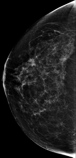
Fig. 99.1 Right craniocaudal (RCC) mammogram.
Related posts:


Stay updated, free articles. Join our Telegram channel

Full access? Get Clinical Tree


