• Denervation changes in muscles distal to entrapment
Top Differential Diagnoses
• Cervical radiculopathy
• Tenosynovitis
• Peripheral nerve sheath tumor
• Thoracic outlet syndrome
Pathology
• Overuse
• Arthritis
• Tumors
• Fractures
• Anatomic variants
Clinical Issues
• Tinel sign: Tingling along course of nerve when nerve tapped at point of entrapment
• Electrodiagnosis standard for diagnosis
Diagnostic Checklist
• Imaging diagnosis challenging
Nerve normally high signal intensity (in carpal tunnel) on FSE T2WI
Nerve normally flattened at level of hook of hamate
• Diagnosis usually made on EMG, not MR
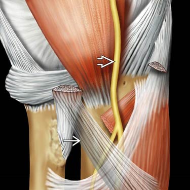
(Left) Graphic of anterior elbow shows median nerve passing anterior to brachialis muscle. It extends between the heads of the pronator teres and beneath the biceps aponeurosis . Entrapment of the nerve in this region is rare compared to carpal tunnel syndrome. It usually presents with numbness with repeated pronation/supination of the forearm.
(Right) Coronal graphic of the carpal tunnel shows the median nerve at the ventral margin of the carpal tunnel, superficial to the flexor tendons.
(Left) Axial T2 FS MR performed at the wrist demonstrates a ganglion cyst in the carpal tunnel, exerting mass effect on the median nerve .
(Right) Axial T1WI MR reveals a lipoma within the carpal tunnel. The flexor tendons are displaced ulnarly, and the median nerve is compressed.
TERMINOLOGY
Synonyms
• Pronator syndrome: Nerve entrapment at pronator teres
• Carpal tunnel syndrome: Nerve entrapment at carpal tunnel
Definitions
• Carpal tunnel: Fibroosseous tunnel at volar aspect of wrist
Contains median nerve and flexor tendons
IMAGING
General Features
• Best diagnostic clue
Increased intrinsic nerve signal on STIR MR
• Location
Elbow or wrist
MR Findings
• Nerve enlargement distal to region of entrapment
• Indistinct nerve fascicles on cross-sectional T2WI
Normal nerve is high signal intensity; no additional increase in signal intensity usually seen
• Perineural enhancement with gadolinium (Gd)
• Increased anterior bowing of flexor retinaculum
Distance from hook of triquetrum to hook of hamate (TH)
Only gold members can continue reading. Log In or Register to continue


 passing anterior to brachialis muscle. It extends between the heads of the pronator teres and beneath the biceps aponeurosis
  . Entrapment of the nerve in this region is rare compared to carpal tunnel syndrome. It usually presents with numbness with repeated pronation/supination of the forearm.
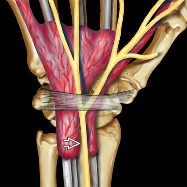
 at the ventral margin of the carpal tunnel, superficial to the flexor tendons.
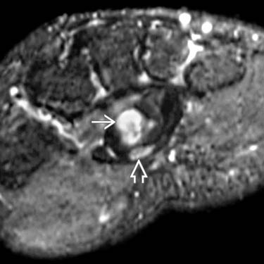
 in the carpal tunnel, exerting mass effect on the median nerve
  .
.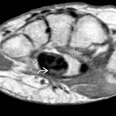
 within the carpal tunnel. The flexor tendons
  are displaced ulnarly, and the median nerve
  is compressed.


