Anatomy
- ◼
The arterial supply of the small bowel is predominantly from the celiac axis and superior mesenteric artery (SMA).
- ◼
The inferior mesenteric artery (IMA) and the internal iliac arteries may become important contributors in the setting of arterial disease.
- ◼
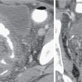
The celiac artery gives origin to the left gastric artery, and then it bifurcates into the splenic and common hepatic arteries ( Fig. 8.1 ).

Fig. 8.1
Selective contrast injection of the celiac artery demonstrates the splenic artery ( short arrow ), common hepatic artery ( single long arrow ), right hepatic artery ( double long arrows ), and gastroduodenal artery ( single arrowhead ).
- ◼
The gastroduodenal artery is a branch of the common hepatic artery (see Fig. 8.1 ) and gives off, among other vessels, the superior pancreaticoduodenal arteries.
- ◼
The SMA arises from the anterior aspect of the aorta at the level of the L1 vertebral body and supplies the duodenum, small bowel, and colon proximal to the splenic flexure ( Fig. 8.2 ).

Fig. 8.2
Selective contrast injection of the superior mesenteric artery ( long arrow ) demonstrates multiple jejunal and ileal branches ( short arrows ), as well as the ileocolic artery ( arrowhead ).
- ◼
The inferior pancreaticoduodenal artery arises from the SMA as the first branch and anastomoses with the superior pancreaticoduodenal artery, forming the pancreaticoduodenal arcades ( Fig. 8.3 ).

Fig. 8.3
Selective contrast injection of the gastroduodenal artery ( white arrowhead ) demonstrates the superior pancreaticoduodenal arteries ( white arrow ), which anastomose with the inferior pancreaticoduodenal arteries ( black arrow ) derived from the superior mesenteric artery ( black arrowhead ).
- ◼
The middle colic artery may arise from the right side of the SMA to supply the transverse colon. It bifurcates into a right branch, which anastomoses with the right colic artery, and a left branch, which anastomoses with the ascending branch of the left colic artery ( Fig. 8.4 ).

Fig. 8.4
Selective contrast injection of the inferior mesenteric artery ( single arrowhead ) demonstrates the ascending ( single short arrow ) and descending ( double short arrows ) branches of the left colic artery. The ascending branch anastomoses with the left branch of the middle colic artery ( single long arrow ), which is derived from the superior mesenteric artery. The marginal artery of Drummond ( double long arrows ) gives off arborizing vasa rectae ( double arrowheads ) to the colon.
- ◼
Multiple jejunal and ileal arteries, as well as the right colic artery supplying the ascending colon arise from the SMA.
- ◼
More distally, the SMA terminates in the ileocolic artery, which supplies the terminal ileum, cecum, and ascending colon.
- ◼
The IMA arises from the left aspect of the aorta at the level of the L3 vertebral body.
- ◼
The IMA divides into the left colic, sigmoid, and superior hemorrhoidal arteries, which supply the descending colon, sigmoid colon, and rectum, respectively. The ascending branch of the left colic artery anastomoses with the left branch of the middle colic artery, a branch of the SMA (see Fig. 8.4 ). The superior hemorrhoidal artery anastomoses with branches of the anterior division of the internal iliac arteries.
- ◼
The marginal artery, defined as the artery closest to and parallel with the mesenteric margin of the intestine, gives off the vasa rectae, which are small vessels that supply the bowel wall. In the colon, this artery is termed the marginal artery of Drummond (see Fig. 8.4 ). The middle colic artery may serve as the marginal artery for much of its distribution.
- ◼
The rich arterial supply of the bowel provides ample opportunity for collateral development in the setting of mesenteric arterial stenosis or occlusion. The superior and inferior pancreaticoduodenal arteries can provide important collaterals between the celiac axis and the SMA (see Fig. 8.3 ). In addition, a persistent fetal arterial communication between the celiac axis and the SMA (often referred to as the arc of Buehler) may exist in up to 2% of patients.
- ◼
Collateral pathways between the SMA and IMA primarily involve communication between the left and middle colic arteries via the arc of Riolan ( Fig. 8.5 ), located centrally in the mesentery, and the marginal artery of Drummond, located peripherally.

Fig. 8.5
Selective contrast injection of the inferior mesenteric artery ( arrowhead ) demonstrates a prominent arc of Riolan ( short arrow ). The arc of Riolan bridges the inferior and superior mesenteric arteries ( long arrow ).
- ◼
The internal iliac arteries also may provide collateral circulation to the principal mesenteric vessels.
- ◼
The superior mesenteric vein (SMV) is usually a single vessel that drains the small intestine and the ascending and transverse colon. Within the mesentery, the SMV courses anteriorly and to the right of the SMA and joins the splenic vein to form the main portal vein.
- ◼
The inferior mesenteric vein (IMV), which drains the left colic, sigmoid, and superior hemorrhoidal veins, usually terminates in the splenic vein or the SMV.
- ◼
Portal-to-portal collaterals may develop in the setting of chronic SMV occlusion; these are often submucosal and are prone to bleeding. Portal-to-systemic collaterals (varices) also may develop in portal hypertension.
Pathophysiology
- ◼
Acute mesenteric ischemia of the small bowel has four major causes ( Table 8.1 ):
- ◼
Arterial embolism
- ◼
Arterial thrombosis
- ◼
Nonocclusive mesenteric ischemia (NOMI)
- ◼
Mesenteric venous thrombosis
Table 8.1
Major Causes of Acute Mesenteric Ischemia
Cause
Onset
Features
SMA embolism
Acute
MI, arrhythmias, ventricular aneurysm, valvular disease, prior embolic event
SMA thrombosis
Acute or acute-on-chronic
Atherosclerotic disease, hypercoagulable state
Nonocclusive ischemia
Acute, failure to thrive
Critical illness, hypotension, MI, sepsis, DIC, vasopressors
Venous thrombosis
Acute or chronic
Recent surgery, hypercoagulable state, OCP Related posts:

Stay updated, free articles. Join our Telegram channel

Full access? Get Clinical Tree


- ◼