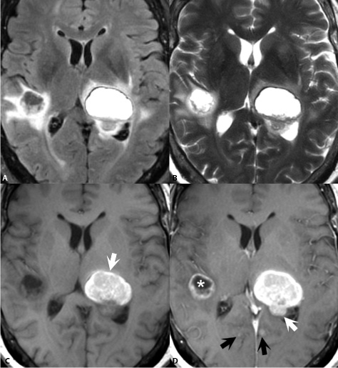
3 Metastatic Brain Disease Metastases comprise about half of all intracranial tumors. Lung cancer followed by breast cancer then melanoma are the most common tumors to metastasize to the brain. Metastatic lesions are confirmed by the presence of multiple intracranial masses (Figs. 3.1A,B,C,D), although this differential also includes less common entities such as multifocal primary brain tumors and abscesses. Solitary brain metastases—seen frequently in breast, uterine, and gastrointestinal (GI) cancer–comprise about half of all brain metastases and pose a diagnostic challenge. Other imaging features suggestive of metastatic brain cancer include well-defined lesions (although pathologically metastases are less well defined than they appear on imaging) and a location at the gray–white matter junction (Figs. 3.1A,B,C,D). The predilection for this region likely relates to the inability of hematogenously spreading tumors to move freely through the smaller vascular structures found there. This hematogenous spread may form the basis for the preponderance of metastatic cancer found in the supratentorial region, reflecting the dominance of the carotid over the vertebrobasilar system. Of unenhanced scan sequences, T2WIs (and in particular fluid attenuated inversion recovery [FLAIR] T2WI, Fig. 3.1A
![]()
Stay updated, free articles. Join our Telegram channel


Full access? Get Clinical Tree


