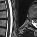
35 Metastatic Disease Of sites of skeletal metastasis, the vertebral column is the most common, and herein lung cancer is the most frequent culprit. Precontrast FSE T1WI is the preferred sequence for the detection of such metastases. As demonstrated in the sagittal images of Fig. 35.1A, infiltration of the vertebral body with tumor results in loss of the high SI of the fatty marrow. Typically, metastatic lesions will demonstrate SI equal to or less than the intervertebral disks on T1WI. This becomes especially important in cases of diffuse metastatic disease, whereby the vertebral body SI may be uniformly abnormal. Only by comparison with the disk SI can diffuse disease be diagnosed. An occasional hemorrhagic metastasis may appear as high SI on T1WI. Unfortunately, FSE T2WI do not typically display most vertebral body metastases well (Fig. 35.1B), due to the relatively high normal marrow SI (see Chapter 48). Even when fat suppression techniques are used, as in Fig. 35.1C
![]()
Stay updated, free articles. Join our Telegram channel


Full access? Get Clinical Tree


