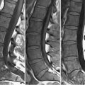
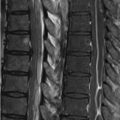
49 Metastatic Disease Metastatic disease at other levels of the spinal column and its effects on the central spinal canal has been previously discussed (see Chapters 35 and 38). The lumbar spine is the second most common location of vertebral metastases (after the thoracic spine), and renal, GI, and prostate cancer demonstrate a predilection for the lumbosacral region. The rationale for the SI changes associated with metastases to the vertebral bodies was described in Chapter 35. Essentially in an adult, there is a majority of fatty compared with hematopoietic marrow, and as such, vertebral bodies demonstrate high SI on T1WI, due to the relatively short T1 of fat. Note that this appearance is largely absent in the vertebral bodies illustrated in Fig. 49.1A. Confluent metastases have resulted in the replacement of high SI marrow fat, resulting in the majority of the lumbar vertebrae, in addition to S1, demonstrating a low SI on T1WI. Higher SI vertebrae or portions thereof as in L5 and S2 reflect normal fatty marrow. FS FSE T2WI (Fig. 49.1B
![]()
Stay updated, free articles. Join our Telegram channel

Full access? Get Clinical Tree


