KEY WORDS
Adenomyosis. Common condition causing pain and heavy periods. Endometrial tissue implants in the myometrium causing uterine enlargement and a variety of sonographic changes. Can be focal or generalized.
Cuff. After hysterectomy, the blind end of the vagina is sutured and may form a fibrous mass, the cuff.
Fibroid (Myoma, Leiomyoma). A benign tumor of the smooth muscle of the uterus. Submucosal—a fibroid bordering on the endometrial cavity. Subserosal—a fibroid bordering on the peritoneal cavity. Intramural—fibroid within the wall of the uterus.
Hematometrocolpos. Metra, uterus; culpa, vagina. Condition presenting at birth or at puberty due to an imperforate hymen. Blood or other fluid accumulates in the vagina and uterus.
Hematocolpos. Obstructed vagina filled with blood. Usually found in teenagers with imperforate hymen or transverse vaginal bands shortly after the onset of menstruation.
Hematometra. Obstructed uterus filled with blood. Usually related to a cervical obstructive process such as cancer of the cervix.
Intramural. Term used to describe a lesion, such as a fibroid, that lies in the wall of the uterus.
Myoma. See Fibroid.
Pedunculated. Term used particularly for fibroids describing a mass that is connected to its site of origin by only a short pedicle.
Submucosal. Term used to describe a process, such as a fibroid, that is located adjacent to the uterine cavity within the uterus.
Subserosal. Term used to describe a lesion such as a fibroid that is on the surface of the uterus.
The Clinical Problem
Masses that appear to involve the uterus are routinely examined with ultrasound for several reasons:
1. It is difficult to assess the ovaries by clinical examination when there is a sizable uterine mass present.
2. Tracking the size of fibroids is most accurately performed with ultrasound. Very rapid growth of an apparent fibroid suggests that the mass might be a leiomyosarcoma.
3. Not all uterine masses are fibroids. Adenomyosis is a common entity enlarging the uterus that is treated in a different fashion.
4. Occasional ovarian masses in the midline are mistaken for masses of uterine origin.
5. Delineating the relationship of the myoma to the endometrial cavity is important in planning surgery.
Anatomy
See Chapter 29.
Technique
ABDOMINAL APPROACH
![]() Uterine masses are often so large that the entire size of the uterus and the size of individual fibroids can only be measured using the abdominal approach.
Uterine masses are often so large that the entire size of the uterus and the size of individual fibroids can only be measured using the abdominal approach.
![]() If the ovaries are visible using the abdominal approach, measure and record their size. They may be located at too high a level to be visible with the endovaginal transducer.
If the ovaries are visible using the abdominal approach, measure and record their size. They may be located at too high a level to be visible with the endovaginal transducer.
VAGINAL APPROACH
![]() You may find that even though fibroids are large, they do not border on the endometrial cavity and are therefore not responsible for vaginal bleeding. Only the improved resolution of the vaginal probe may show this relationship if the endometrial cavity is difficult to see.
You may find that even though fibroids are large, they do not border on the endometrial cavity and are therefore not responsible for vaginal bleeding. Only the improved resolution of the vaginal probe may show this relationship if the endometrial cavity is difficult to see.
![]() When an intracavitary mass is suspected in the presence of fibroids, use a balloon catheter with a saline infusion study; the fibroids are solid and not easily displaced by the intracavitary fluid unless a balloon catheter is used.
When an intracavitary mass is suspected in the presence of fibroids, use a balloon catheter with a saline infusion study; the fibroids are solid and not easily displaced by the intracavitary fluid unless a balloon catheter is used.
Clinical
UTERINE MASSES
Solid Uterine Masses
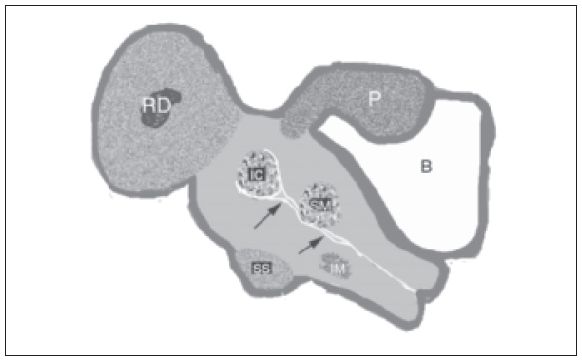
Fibroids (leiomyomas). Fibroids represent an overgrowth of uterine smooth muscle that forms a tumor. Leiomyoma is the benign form, and leiomyosarcoma is the rare malignant form. Fibroids are the most common tumors in women and are present in 40% of women aged more than 40 years. They usually grow progressively during the menstrual years but may shrink after menopause. Common symptoms are heavy, prolonged periods; infertility; and pelvic pain. They may be intracavitary, submucosal, intramural, subserosal, or pedunculated (Fig. 31-1).
Sonographically, the features are as follows:
1. An enlarged uterus, usually with a lobulated contour that may indent the bladder (Fig. 31-2). If the bladder volume is small, document the size. Frequency is a common complication of fibroids because they reduce bladder capacity.
2. Focal ovoid or circular masses within the uterus. These masses may have a similar echogenicity to the remainder of the uterus, but tissue within is organized in a whirled (circular) fashion (Fig. 31-3). Blood vessels form a rim around the fibroid, whereas with other entities they may look similar, such as in focal adenomyosis the blood vessels traverse the lesion.

Figure 31-1. ![]() Fibroids may be located in several different sites. Subserosal fibroids (SS) lie on the edge of the uterus and may indent the bladder. They are almost always asymptomatic. Intramural fibroids (IM) lie in the center of the myometrium (the muscular component of the uterus). If they do not distort the cavity, they are usually asymptomatic. Submucosal fibroids (SM) lie on the edge of the endometrium. They often cause menstrual cramping and bleeding. Intracavity fibroids (IC) almost always cause cramping and bleeding. Pedunculated fibroids (P) are usually asymptomatic, but in this diagram, the fundal pedunculated fibroid has an echopenic center because it has undergone red degeneration (RD). This painful condition usually occurs in pregnancy.
Fibroids may be located in several different sites. Subserosal fibroids (SS) lie on the edge of the uterus and may indent the bladder. They are almost always asymptomatic. Intramural fibroids (IM) lie in the center of the myometrium (the muscular component of the uterus). If they do not distort the cavity, they are usually asymptomatic. Submucosal fibroids (SM) lie on the edge of the endometrium. They often cause menstrual cramping and bleeding. Intracavity fibroids (IC) almost always cause cramping and bleeding. Pedunculated fibroids (P) are usually asymptomatic, but in this diagram, the fundal pedunculated fibroid has an echopenic center because it has undergone red degeneration (RD). This painful condition usually occurs in pregnancy.
Stay updated, free articles. Join our Telegram channel

Full access? Get Clinical Tree


