Fig. 34.1
A 2-month-old girl presented with the swelling of bilateral forearm. (a) Typical periosteal new bone formation (arrows) of mandible. (b, c) Periosteal new bone formation (arrows) of radius and ulna
34.3.2 Periosteal New Bone Formation
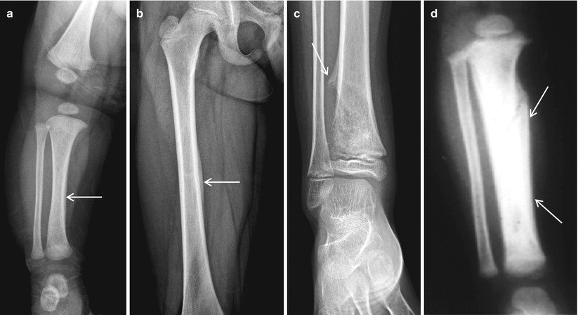
Fig. 34.2
Periosteal new bone formation. (a) Normal infant. (b) Single layer of periosteal new bone formation (arrow). This case is associated with incomplete fracture. (c) Typical Codman’s triangle in the malignant bone tumor. This patient had an osteosarcoma. (d) Extensive periosteal new bone formation in the battered child syndrome
34.3.3 Bone Infarction
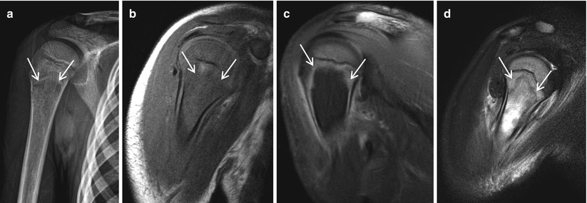
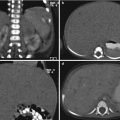
Fig. 34.3
A 6-year-old boy presented with the pain of right humerus. He had chemotherapy due to leukemia. (a) Osteolytic lesion (arrows) in humerus. (b) Low-signal-intensity lesion in the T1-weighted MR image. (c) Nonenhancing lesion (arrows) in T1-weighted MR image with the enhancement of gadolinium. (d) High-signal-intensity lesion (arrows) in the T2-weighted MR image
34.3.4 Kohler Disease
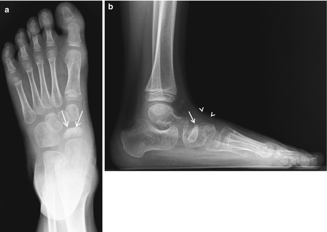
Fig. 34.4
A 6-year-old boy presented with foot pain. (a, b) Irregularity and sclerosis of the navicular bone (arrows) with soft tissue swelling (arrowheads)
34.3.5 Freiberg Disease
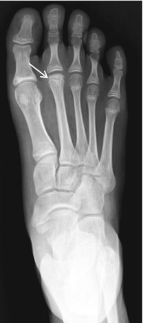
Fig. 34.5
A 13-year-old girl presented with foot pain. Note the flattening and sclerosis of secondary metatarsal bone (arrow)
34.3.6 Sever Disease
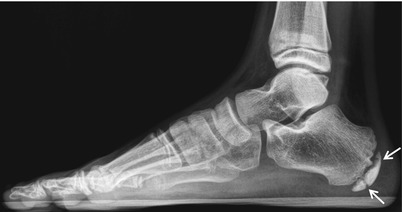
Fig. 34.6
An 11-year-old boy presented with heel pain, demonstrating the sclerosis and fragmentation of calcaneus (arrows)
34.3.7 Idiopathic Juvenile Osteoporosis
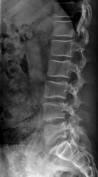
Fig. 34.7
A 13-year-old boy presented with the back pain. Note the biconcave appearance of vertebral bodies with diffusely decreased bone density
34.3.8 Hypothyroidism
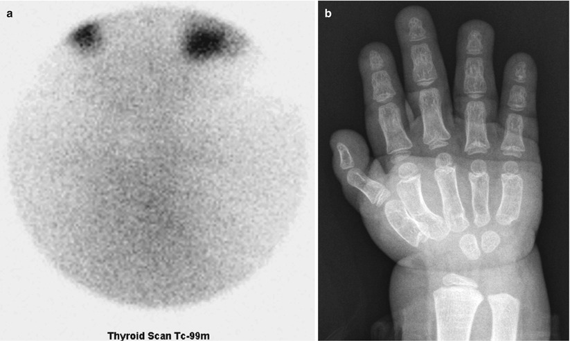
Fig. 34.8
A 4-year-old girl presented with developmental delay. (a) No definite uptake of thyroid gland on the thyroid scan. (b) Note the delay of bone age
34.3.9 Hypopituitarism and Growth Hormone Deficiency
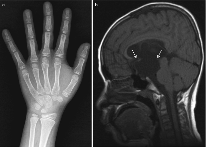
Fig. 34.9
A 12-year-old boy presented with suprasellar arachnoid cyst. (a) Note the delay of bone age. (b) Suprasellar arachnoid cyst (arrows) is demonstrated
34.3.10 Hyperparathyroidism
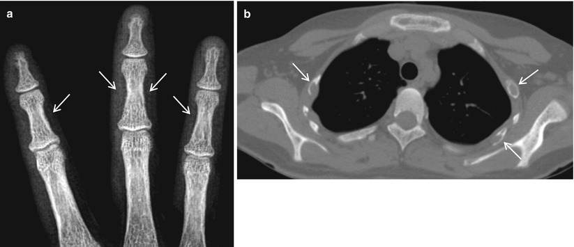
Fig. 34.10
A 16-year-old girl presented with multiple bone masses. (a) Typical subperiosteal bone resorption (arrows) of the middle phalanges. (b) Multiple osteolytic masses (arrows) in ribs, which are the pathologically proven brown tumor
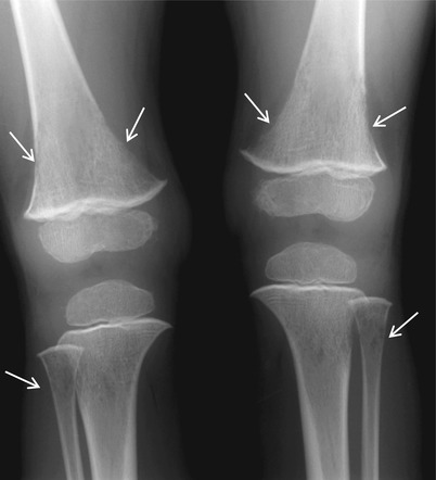
Fig. 34.11
A 2-year-old boy presented with knee pain. Note the subperiosteal resorption (arrows) around the bilateral knees
34.3.11 Pseudohypoparathyroidism
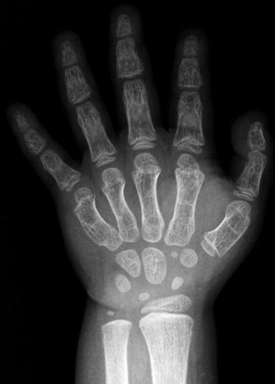
Fig. 34.12


A 4-year-old girl presented with the short length of finger. Characteristic shortening of the fourth and fifth metatarsal bones with slightly ballooned bones
Related posts:


Stay updated, free articles. Join our Telegram channel
Full access? Get Clinical Tree
