Fig. 31.1
T-tube (Printed with permission from Hood Laboratory)

Fig. 31.2
Variation of T-tubes (Printed with permission from Hood Laboratory)
The T-tube size ranges from the pediatric 6.0 mm diameter to 16 mm accommodating different airway sizes in both small children and adults. It minimizes the risk of migration which is especially important given its proximity to the vocal cords. In cases of stenosis or surgical resection of high tracheal disease, the proximal portion of the T-tube can be placed above the vocal cords. The wide variety of options provides either a clear or radiopaque T-tube. For the patient who has a proximal subglottic stenosis, the proximal T-tube limb is tapered to accommodate this while providing a more viable airway. The length of the individual arms allow for patient specific customization. Measurements can be provided to the manufacturer and a custom tube can be made very efficiently. However, the physician is also free to customize the standard tube onsite at the time of the procedure. It is very important when shortening the limbs the intraluminal ends must be smoothed and beveled to reduce formation of granulation tissue. This can be done by cutting the tube with a scalpel blade and smoothing the edges with sandpaper. There is a customizing kit that can be ordered by the manufacturer. To properly size both the diameter and length of the T-tube, CT scan imaging of the trachea with virtual images, 3D reconstruction, coronal, axial, and sagittal views are very helpful. Bronchoscopic visualization preprocedure is also helpful to assist with measurement confirmation. It is important to be mindful if the proximal disease permits, to avoid allowing the proximal vertical limb to be at or < 5 mm from the true vocal cords. It is also important to assure that the internal airway limbs of the stent cover the pathologic site and that all limbs fully open upon placement. If there is notable persistent infolding of any of the limbs after placement, then this likely indicates that the stent diameter is too large and must be replaced with a smaller diameter tube. Ideally, the fit would be flush to the mucosa limiting any sliding movement of the tube against the dynamic tracheal walls.
Indications
Initially developed for use in the setting of providing support for a reconstructed trachea and tracheal resection and anastomosis, and acute tracheal injury, the indication has expanded to cover other etiologies of upper airway obstruction requiring structural support. The indication can be divided based on the etiology of airway disease: neoplastic and nonneoplastic.
Neoplastic Etiology
This category includes airway obstruction secondary to either primary tracheal tumors or metastatic diseases of the airway. Metastatic cancers that can involve the trachea such as the esophagus, as well as those mediastinal tumors that may result in extrinsic airway compression such as thyroid or lymphoma, may create comprise to airway function.
Nonneoplastic Etiology
This category of disease includes postanastomotic stricture and postintubation stenosis. Less common reasons include stenosis associated with tuberculosis, amyloidosis, sarcoidosis, and postradiation stricture. Aneurysms of the vascular structure surrounding the airway or congenital abnormalities can cause extrinsic compression and airway obstruction. It must be noted that surgical correction of nonneoplastic tracheal stenosis is the gold standard, and stents such as the T-tube should be used in surgically uncorrectable cases either due to prohibitive patient surgical risk factors and technical difficulties or as a bridge to definitive surgical correction. However, there have been many cases where the T-tube has been left in place for prolonged periods of time up to 16 years without any significant complications or patient intolerance. A T-tube due to its anchored placement may be more optimal than a standard cylindrical silicone tracheal stent based on disease that may be more proximal in the trachea with the possibility of tracheal stent migration to the vocal cords. In those patients who require more frequent pulmonary toilet with a suboptimal cough, the T-tube provides a much needed access into the airway for suctioning that would not be possible with a tracheal stent alone. A T-tube may be more comfortable and more cosmetically appealing to patients.
In respect to the surgical management of tracheal disease, the T-tube can be used as an adjunct to surgical management:
1.
Temporary airway support prior to definitive surgical resection.
2.
Compliment high tracheal and subglottic resection postsurgery: the T-tube is placed at the time of the surgery and left in place for prolonged period, allowing appropriate airway remodeling and preventing postsurgical stenosis.
3.
Failed primary tracheal resection: in patients who develop restenosis postsurgically or partial separation of anastomosis can benefit from prolonged T-tube use.
4.
Primary definite therapy with T-tube if surgically unfeasible: the T-tube can be placed either indefinitely or for a prolonged period to allow airway remodeling.
Contraindications
In patients likely to require prolonged positive pressure ventilation in the immediate future, the T-tube would not be the recommended option due to the proximal air leak from the proximal tracheal limb of T-tube. This will be discussed further in detail below. Placement of a Montgomery T-tube must be cautiously considered in patients with significant thick respiratory secretions due to a higher incidence of possible stent occlusion from mucus impaction. One case series showed a 9.3% complication rate secondary to secretion retention, requiring intervention such as bronchoscopy. Caution should always be used when the tube inner diameters are small, as seen in children due to the risk of airway obstruction from dried secretions. Meticulous care in sizing, position, and cleaning is required.
In cases of the proximal vertical limb of the T-tube extending above the vocal cords preventing full glottis closure, there is an increased risk of aspiration. Patients who already have a clear history of aspiration should not undergo T-tube placement.
Insertion and Removal
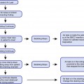
The presence of a tracheostomy opening is a mandatory prerequisite for placement of the T-tube. The original insertion method described by Dr. Montgomery with some modifications is as follows: the airway is secured with a rigid tracheoscopy tube with jet ventilation applied via a working side channel of the tracheoscope, and then using a hemostat, grasp the inferior limb of the vertical tube after folding it upon itself. This is inserted through the tracheostomy opening inferiorly. The hemostat is released and then used to grab the proximal vertical limb next inserting it superiorly. The external limb is pulled gently to allow the other limbs to open up and be directed appropriately (Fig. 31.3).
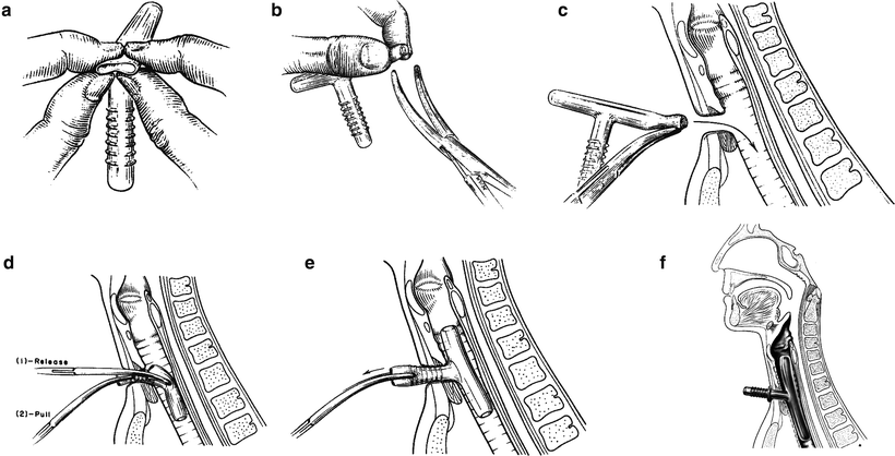

Fig. 31.3
Demonstration of one method of placing a T-tube (Printed with permission from Boston Medical)
There have been many new approaches and modifications to the original method described. One popular method suggested by Cooper et al. in 1981 involves using the rigid bronchoscope and an umbilical tape threaded through the horizontal limb, proximal limb of the T-tube, then through the tracheostomy opening and exit through the rigid bronchoscope at the vocal cords. By inserting the inferior limb then pulling on the umbilical cord, this allows quick insertion and alignment of the T-tube. Subsequently, many authors published variations of this method using the umbilical tape and either the rigid bronchoscope or endotracheal tube.
Other interesting methods suggested by some authors involve loading the T-tube onto the endotracheal tube prior to insertion. A size 6.0 mm endotracheal tube in the adult can either be inserted through the horizontal limb into the distal vertical limb or the proximal vertical limb of the T-tube then inserted into the trachea and exited inserted into the trachea and exited out through the tracheostomy site, then pulled back into the tracheostomy site.
Stay updated, free articles. Join our Telegram channel

Full access? Get Clinical Tree


