Morton Neuroma Syndrome
Anatomic Considerations
In a manner analogous to that of the digital nerves of the hand, the digital nerves of the foot travel through the intrametatarsal space to innervate each toe. The plantar digital nerves, which are derived from the posterior tibial nerve, provide sensory innervation to the major portion of the plantar surface (Fig. 22.1). These nerves are subject to entrapment and resultant development of perineural fibrosis and degeneration resulting in the clinical syndrome known as Morton neuroma (Fig. 22.2). The dorsal aspect of the foot is innervated by terminal branches of the deep and superficial peroneal nerves. The overlap of the innervation of these nerves may be considerable.
 FIGURE 22.1 Anatomy of the metatarsophalangeal and digital nerves. |
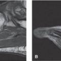
 FIGURE 22.2 Patients suffering from Morton neuroma present with the complaint of pain in the plantar surface with associated dysesthesias radiating into the adjacent toes. |
Clinical Correlates
Morton neuroma, which was first described by Thomas Morton in 1876 refers to a constellation of symptoms including tenderness and burning pain in the plantar surface of the forefoot, with associated painful dysesthesias into the affected two toes. This pain syndrome is thought to be caused by perineural fibrosis of the interdigital nerves (Fig. 22.3). There is often coexistent intermetatarsal bursitis as the pathogenesis of both pathologic conditions is similar (Fig. 22.4). Although the nerves between the third and fourth toes most often are affected, the second and third toes and, rarely, the fourth and fifth toes can be affected.
Most patients with Morton neuroma present with the complaint of pain in the plantar surface with associated dysesthesias radiating into the adjacent toes. Patients commonly complain that it feels like they are walking with a stone caught in their shoe. Walking, standing, or wearing tight shoes makes the pain worse, with rest and heat providing some relief. The pain is constant and is characterized as aching and may interfere with sleep.
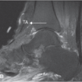
 FIGURE 22.3 Morton neuroma. Axial T1-weighted (A) and postcontrast fat-suppressed T1-weighted (B) images demonstrating neuromas between the second and third and third and fourth metatarsal bases. C: Illustration of the location of Morton neuromas and intermetatarsal bursae with relation to the transverse metatarsal ligament. (From Berquist TH. Imaging of the Foot and Ankle. 3rd ed. Philadelphia, PA: Lippincott Williams & Wilkins; 2011:200.) |
On physical examination, the pain associated with Morton neuroma can be reproduced by performing the Mulder maneuver. This is accomplished by firmly squeezing the two metatarsal heads together with one hand while placing firm pressure on the interdigital space with the other hand (Fig. 22.5). A palpable click may also be appreciated as the neuroma is forced from between the metatarsals. The patient
with Morton neuroma often exhibits an antalgic gait in an effort to reduce weight bearing during walking.
with Morton neuroma often exhibits an antalgic gait in an effort to reduce weight bearing during walking.
 FIGURE 22.4 Morton neuroma change with an associated small effusion in the intermetatarsal bursa. Anechoic fluid (arrow) is seen within the bursa at the proximal dorsal aspect of typical Morton web space change (area between X’s). Distal is to the left of the image. (From Read JW, Noakes JB, Kerr D, et al. Morton’s metatarsalgia: sonographic findings and correlated histopathology. Foot Ankle Int 1999;20(3):153–161.) |
 FIGURE 22.5 Mulder maneuver is accomplished by firmly squeezing the two metatarsal heads together with one hand while placing firm pressure on the interdigital space with the other hand. (From Berg D, Worzala K. Atlas of Adult Physical Diagnosis. Philadelphia, PA: Lippincott Williams & Wilkins; 2006.) |
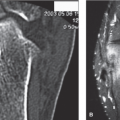
 FIGURE 22.6 Axial T1-weighted (A), T2-weighted (B), and postcontrast enhanced fat-suppressed T1-weighted (C) images demonstrate a Morton neuroma between the third and fourth metatarsals that extends dorsally (arrow). Note the mixed signal intensity on the T2-weighted image (B) and intense enhancement (C). (From Berquist TH. Imaging of the Foot and Ankle. 3rd ed. Philadelphia, PA: Lippincott Williams & Wilkins; 2011:426.)
Related posts: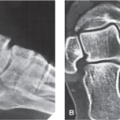 Arthritis and Other Abnormalities of the Subtalar Joint Arthritis and Other Abnormalities of the Subtalar Joint
 Arthritis and Other Abnormalities of the Ankle Joint Arthritis and Other Abnormalities of the Ankle Joint
 Anterior Tarsal Tunnel Syndrome and Other Abnormalities of the Deep Peroneal Nerve Anterior Tarsal Tunnel Syndrome and Other Abnormalities of the Deep Peroneal Nerve
 Retrocalcaneal and Achilles Bursitis Retrocalcaneal and Achilles Bursitis
 Calcaneal Spurs Calcaneal Spurs
 Plantar Fasciitis and Other Abnormalities of the Plantar Fascia Plantar Fasciitis and Other Abnormalities of the Plantar Fascia
Stay updated, free articles. Join our Telegram channel
Full access? Get Clinical Tree
 Get Clinical Tree app for offline access

|