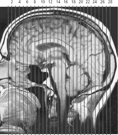
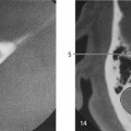
Scout views of coronal MR series
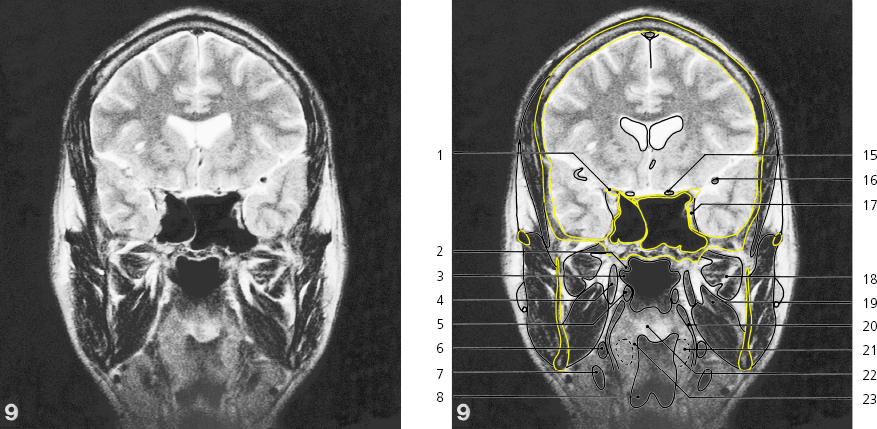
Lines #1–28 indicate positions of coronal sections in the following MR series.Brain , coronal MR
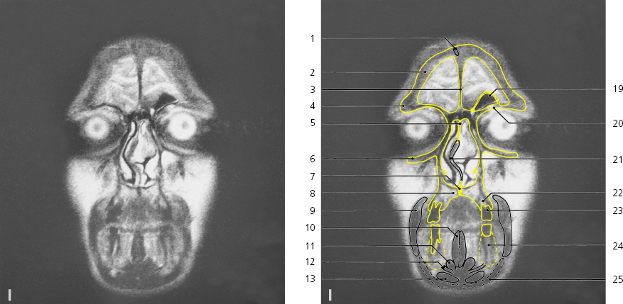
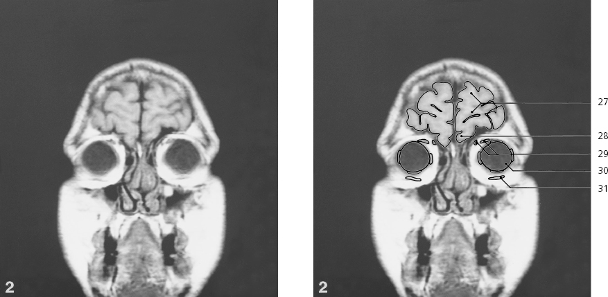
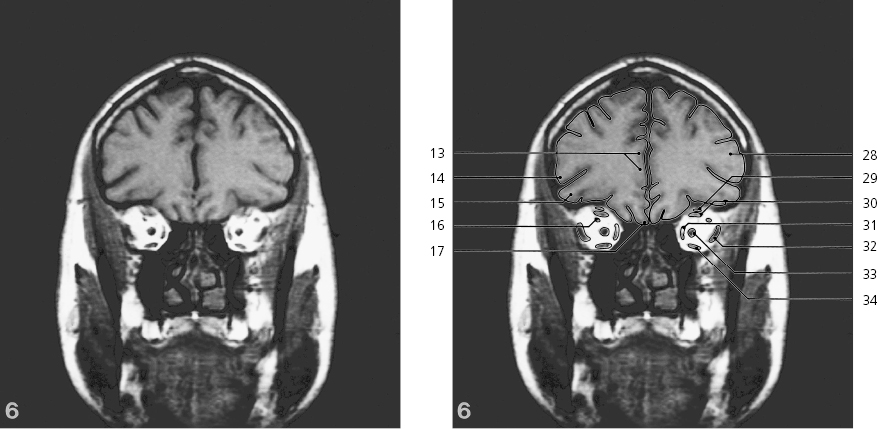
Scout view on previous pageDiploic vein → Frontal bone, squamous part → Frontal crest Supraorbital margin of frontal bone → Perpendicular plate of ethmoidal bone → Infraorbital margin of maxilla → Median palatine suture → Hard palate → Buccinator → Genioglossus → Geniohyoideus → Mylohyoideus → Digastricus, anterior belly → Frontal pole of brain Obliquus superior muscle in trochlea → Lacrimal sac Lens Palpebral fissure Frontal sinus Orbital part of frontal bone → Cartilaginous part of nasal septum → Alveolar part of maxilla → Second upper premolar tooth Alveolar part of mandible → Platysma → Eyelids Brain , coronal MR
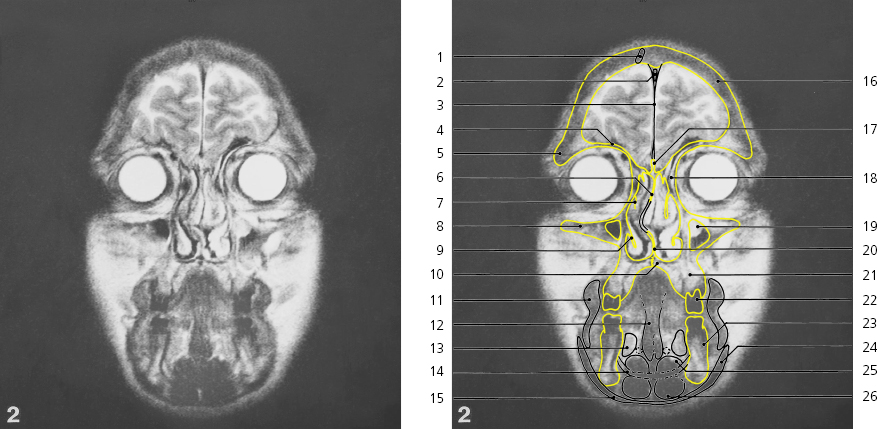
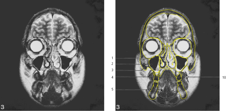
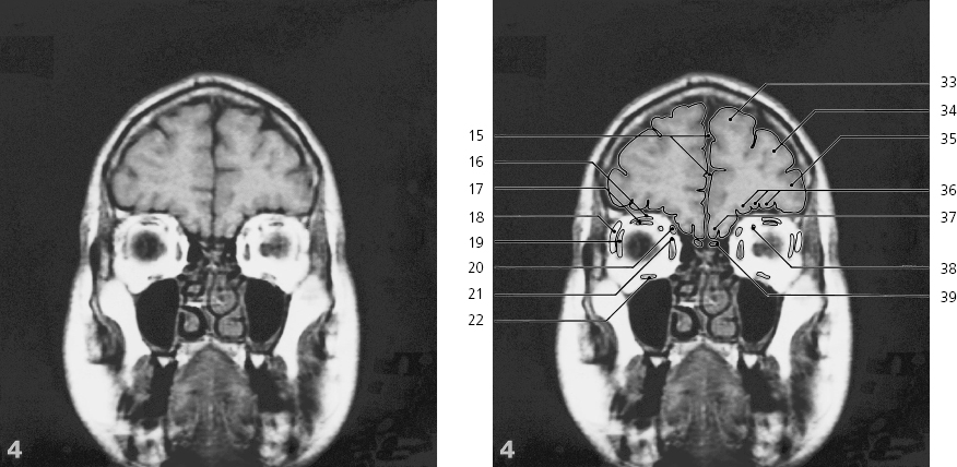
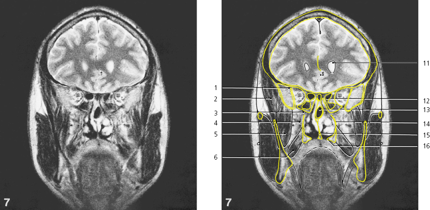
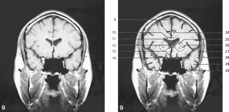
Scout view on page 276Diploic vein ↔ Superior sagittal sinus → Falx cerebri → Orbital part of frontal bone ↔ Supra-orbital margin of frontal bone ← Perpendicular plate of ethmoidal bone ↔ Middle concha → Infra-orbital margin of maxilla ← Inferior choncha ↔ Hard palate ↔ Buccinator ↔ Genioglossus ↔ Sublingual gland → Mylohyoideus ↔ Platysma ↔ Squamous part of frontal bone ↔ Crista galli → Ethmoidal air cells → Maxillary sinus → Vomer → Alveolar part of maxilla ↔ First upper molar tooth Alveolar part of mandibula ↔ Platysma ↔ Geniohyoideus ↔ Digastricus, anterior belly ↔ Frontal lobe ↔ Straight gyrus → Obliquus superior muscle ↔ Eyeball ↔ Obliquus inferior muscle → Brain , coronal MR
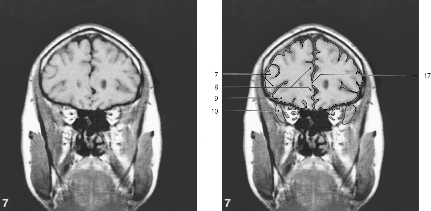
Scout view on page 276Orbital plate of maxilla ↔ Maxillary sinus ↔(with oedematous mucous membrane) Body of zygomatic bone → Tongue ↔ Submandibular duct → Longitudinal fissure of brain ↔ Straight gyrus ↔ Orbital gyri → Lacrimal gland → Facial artery/vein Obliquus superior ↔ Levator palpebrae superioris ↔ Rectus superior ↔ Rectus medialis ↔ Rectus lateralis ↔ Rectus inferior ↔ Obliquus inferior ← Brain , coronal MR
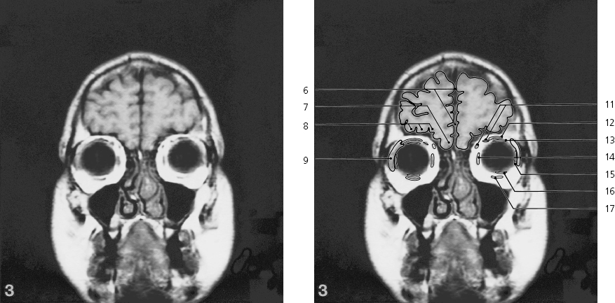
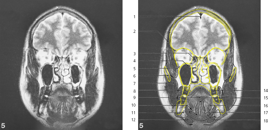
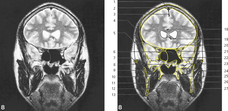
Scout view on page 276Superior sagittal sinus ↔ Squamous part of frontal bone ↔ Orbital part of frontal bone ↔ Temporal fascia → Temporalis muscle → Frontal process of zygomatic bone Greater wing of sphenoid bone in lateral wall of orbita → Body of zygomatic bone ← Masseter Parotid duct → Buccinator ↔ Sublingual gland ← Submandibular duct surrounded by deep part of gland ↔ Platysma ↔ Longitudinal fissure of brain ↔ Levator palpebrae superioris ↔ Rectus superior ↔ Lacrimal gland ← Rectus lateralis ↔ Obliquus superior ↔ Rectus medialis ↔ Rectus inferior ↔ Crista galli ↔ Nasal septum ↔ Middle concha ↔ Inferior concha ↔ Hard palate ↔ Second upper molar tooth Genioglossus ↔ Mylohyoideus ↔ Geniohyoideus ↔ Digastricus, anterior belly ↔ Superior frontal gyrus → Middle frontal gyrus → Inferior frontal gyrus → Orbital gyri ↔ Straight gyrus ↔ Ophthalmic artery → Olfactory bulb Brain , coronal MR
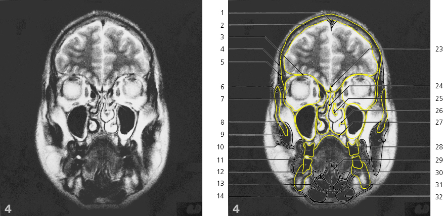
Scout view on page 276Falx cerebri ↔ Temporalis muscle ↔ Ethmoid sinus ↔ Maxillary sinus ↔ Infra-orbital fissure → Zygomatic arch → Masseter ↔ Buccinator ↔ Tongue ↔ Submandibular duct ← Submandibular gland ← Digastricus, anterior belly ↔ Optic nerve → Third upper molar tooth Lingual septum Hyoglossus → Mylohyoideus ↔ Geniohyoideus ↔ Outer lamina of frontal bone Diploë of frontal bone Inner lamina of frontal bone Brain , coronal MR
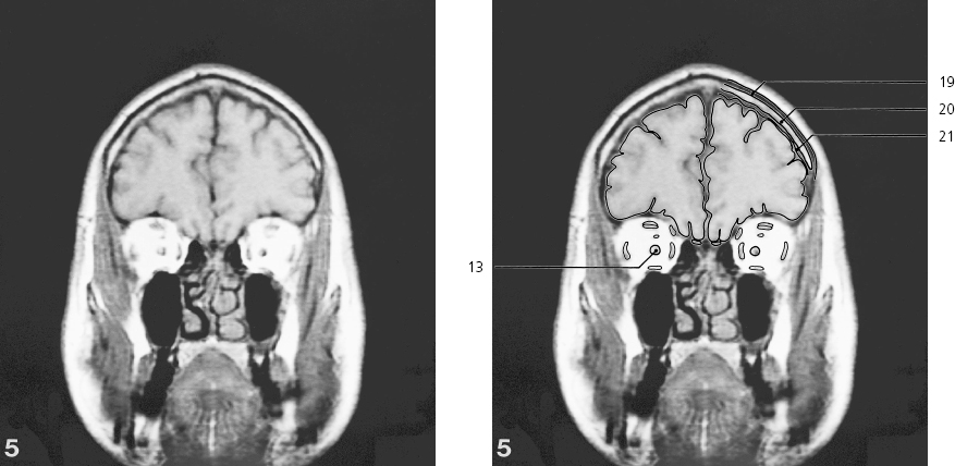
Scout view on page 276Superior sagittal sinus ↔ Falx cerebri ↔ Squamous part of frontal bone ↔ Orbital part of frontal bone ↔ Temporalis muscle ↔ Temporal fascia ↔ Zygomatic arch ↔ Accessory parotid gland Parotid duct ↔ Masseter ↔ Ramus of mandible → Platysma ← Cingulate gyrus → Middle frontal gyrus ↔ Inferior frontal gyrus ↔ Ophthalmic artery ← Straight gyrus ↔ Ethmoid sinus ↔ Greater wing of sphenoid bone ↔ Infra-orbital fissure ↔ Maxillary sinus ←(with oedematous mucous membrane) Buccinator ← Tongue ↔ Mylohyoideus ↔ Hyoglossus ↔ Geniohyoideus ← Digastricus, anterior belly ← Middle frontal gyrus ↔ Levator palpebrae superioris ↔ Rectus superior ↔ Rectus medialis ↔ Rectus lateralis ↔ Optic nerve ↔ Rectus inferior ↔ Brain , coronal MR
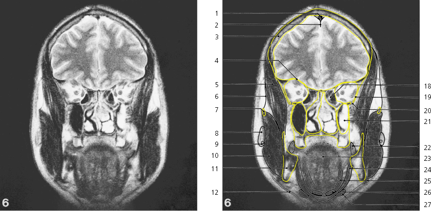
Scout view on page 276Superior orbital fissure ↔ Temporalis muscle ↔ Foramen sphenopalatinum Pterygopalatine fossa Pterygoideus lateralis muscle → Pterygoideus medialis muscle → Middle frontal gyrus ↔ Cingulate gyrus ↔ Inferior frontal gyrus ↔ Anterior pole of temporal lobe → Lateral ventricle, frontal horn → Ethmoidal sinus ← Sphenoidal sinus → Perpendicular plate of palatine bone Vomer Pterygoid process Corpus callosum, genu → Brain , coronal MR
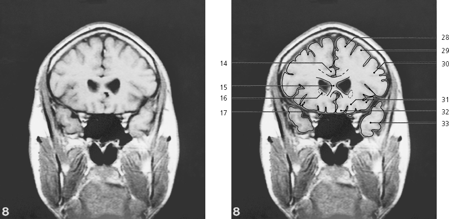
Scout view on page 276Superior sagittal sinus ↔ Squamous part of frontal bone ↔ Falx cerebri ↔ Temporalis muscle ↔ Temporal fascia ↔ Sphenoidal sinus ↔ Zygomatic arch ↔ Vomer, attachment on sphenoidal bone ← Coronoid process of mandible → Pterygoid hamulus Ramus of mandible → Soft palate → Tongue ← Cingulate gyrus ↔ Corpus callosum, genu ↔ Caudate nucleus, head → Optic nerve ↔ Pericallosal artery ↔ Anterior cerebral artery → Optic canal Apex of orbita Foramen rotundum with maxillary nerve Lateral pterygoid muscle ↔ Masseter ↔ Lateral lamina of pterygoid process Medial lamina of pterygoid process ← Pterygoideus medialis muscle ↔ Superior frontal gyrus ↔ Middle frontal gyrus ↔ Inferior frontal gyrus ↔ Gyri orbitales ← Straight gyrus ↔ Temporal lobe ↔ Brain , coronal MR
Only gold members can continue reading.
Log In or
Register to continue
Stay updated, free articles. Join our Telegram channel
Join